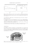
JOURNAL OF COSMETIC SCIENCE 316 The structure of mature HFs is composed of ectoderm- or mesoderm-derived tissue com- partments that are arranged in the shape of an inverted-wine glass with many layers like in an onion. The distal compartment of the HF, close to the skin surface, is the infun- dibulum, which continues into the isthmus, delimited by the insertion of the sebaceous gland and arrector pili muscle, and the suprabulbar and bulbar areas (1,8,9). The isthmus area contains the so-called “stem cell niche,” the bulge, where the multipotent epithelial HF stem cells responsible for the constant renewal of the HFs during the hair cycle reside (10–13). The hair bulb is called the “HS machinery” because it is the HF part where hair matrix (HM) keratinocytes, melanocytes, and highly specialized inductive fi broblasts in the der- mal papilla interact with each other to make the hair fi ber (1,8,9). The HF during its growth phase (anagen) is composed of more than 20 different cell populations. The HF’s epithelial compartments, from the outermost to the innermost layer, are the outer root sheath (ORS), the inner root sheath (IRS), and the hair fi ber. The ORS represents the continuation of the epidermal basal layer. The IRS is further divided into four parts, the companion layer, Henle’s layer, Huxley’s layer, and IRS cuticle and reaches the insertion of the sebaceous gland duct. The HS is constituted by the cuticle, cortex and, only in some cases, the medulla. A substantial basement membrane surrounds the ORS, which in turn is enclosed by a mesenchyme-derived layer, the perifollicular con- nective tissue sheath (1,8,9,14,15). The HF undergoes life-long process of cyclic transformations (1,8,16–18). The hair fi ber is only produced during the HF growth phase, called anagen, which is proportional to the size of the dermal papilla, dictating the size, shape, and length of the hair (19). The growth phase is followed by an apoptosis-driven regression phase called catagen and by a relative stage of rest (telogen). Approximately 100,000 terminal HFs are found in the human scalp, which produce dead hair fi bers that are typically long but can vary in shape, color, and physical behavior. The growth of the hair fi bers in the human scalp is reported to be around 0.3–0.5 mm per day (20). During anagen, the HS and IRS are produced by highly proliferating germinative HM keratinocytes generated by stem cells that originated from the bulge and migrated to the hair bulb during early anagen (10–13). These cells lose their proliferative activity after a critical anatomically defi ned level in the follicle structure (called as the Auber line), and start to differentiate into trichocytes of all HF epithelial cell layers with the exception of the ORS (9,14,21). The mechanisms that are involved in proliferation–differentiation switch of the HM keratinocytes remain largely unknown. The HS production is associ- ated with the initiation of the hair-specifi c keratin gene expression program (13,14,22,23). The mystery of what occurs at the border between the germinative and precortical HM remains one of the major fascinating open questions in the fi eld. It has recently been shown that Cyclin-dependent kinase (CDK) interacting protein/kinase inhibitory protein (CIP/KIP) family members regulating cell cycle progression/arrest, differentiation, and endoreplication are involved in this process (13). Other factors have also been shown to be implicated in the switch between HM keratinocytes proliferation and differentiation, such as the distance from the dermal papilla, which supply a gradient of growth factors (14), as well as miRNAs and other epigenetic regulators (24). The HS is composed of dead (fully differentiated) keratinocytes (cortical cells) containing mainly proteins. Although the cuticle comprises 6–11 overlapping cell sheaths with an

BRIDGING THE “DEAD HAIR”—“LIVE FOLLICLE” 317 exposed edge, cells in the cortex become elongated and the hair keratins are organized into macrofi brils (15,25,26). These are complex structures containing intermediate fi la- ments embedded in a matrix composed of keratin-associated proteins (KAPs). The inter- mediate fi laments are collections of heterodimers of hair keratin proteins. Macrofi bril structures are stabilized by cross-links established by a range of disulphide bonds, ionic bonds and hydrogen bonds (25–27). The array of hair keratins and KAPs expressed in the HS and other HF compartments have been very well characterized (14,22,23,28– 30). Although these clearly determine the structure and strength of the HS, researchers have failed to understand how the modulation of HM keratinocyte activities correlates with hair keratins and KAP’s expression and infl uences HS properties. Some attempts have been made by “live follicle” researchers, who have shown that precortical hair kera- tins can be regulated by selected endogenous and exogenous factors ex vivo, using human HF organ culture (31,32). Unfortunately, methodological limitations prevented these researchers from translating their fi ndings to changes in HS quality. In addition, de- creased expression of selected hair keratins and KAPs is correlated with hair ageing (33), which is also characterized by thinning of the HS (34). Ultimately, the changes occurring in the “live follicle” contribute to alterations in char- acter of “dead hair” structure and quality however, research within the two HF worlds remains separated. This is quite surprising considering that these two worlds share the hair care market and try to satisfy common consumers. The “live follicle” and “dead hair” researchers have clearly different scientifi c interests and different backgrounds biologists are unraveling the secret life of the “live follicle,” whereas physicists and chemists are involved in learning about the material properties of the “dead hair.” Indeed, an intense literature search is required to reveal the aspects of the “live follicle” that affect the “dead hair” structure and quality. Most of such studies come from the wool industry, for which obviously wool hair fi ber quality is more important than hair fi ber length and, therefore, anagen maintenance. For example, an elegant study by Bond et al. (35,36) showed that ex vivo treatment of wool adult anagen HFs with fi broblast growth factors 1 and 2, whose receptors are expressed in the living HFs, has an impact on protein synthesis for the “dead hair” but did not alter the cells of the “live” HM. Although the authors have not analyzed the hair fi ber quality, they suggested that these changes likely affect tensile strength. In humans, some information can be extrapolated from genetic hair conditions. It is clear that anything that interferes with the production of “dead hair” shaft keratins during keratinocyte differentiation in the “live follicle” results in decrease in HS thickness and integrity, as seen in monilethrix (5,23,37). Regulation of the production of trichohyalin (THH) or its solubility contributes to HS abnormalities. Also, genetic mutations of genes encoding for THH or enzymes involved in THH post-translational modifi cation result in the condition called uncombable hair syndrome, manifested by a production of silvery- blond or yellowish-colored disorderly hairs, which are diffi cult to comb (38). Hair disor- ders characterized by abnormal HS curvature have been linked to changes in the expression of IRS keratins (3,5). Patients with defi ciency in Adenosine triphosphate (ATP)7A (or Menkes’ ATPase), which is involved in copper traffi cking, feature hypopigmented brittle hairs, also called kinky hairs (39,40). Mutation in genes encoding for a protein involved in the cell cycle, namely, M phase–specifi c Serine/threonine-protein kinase (PLK1), causes trichothiodystrophy, a syndrome in which hairs are brittle because of defi ciency in sulfur, a fundamental component for intermediate fi lament cross-linking in the HS (41). We have
Purchased for the exclusive use of nofirst nolast (unknown)
From: SCC Media Library & Resource Center (library.scconline.org)