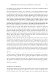
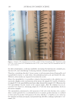
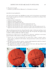
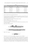
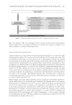
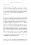
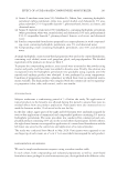
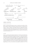
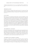
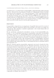
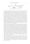
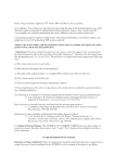
DISINFECTION OF REUSABLE MAKE-UP APPLICATORS 165 6. On e protective mask, 7. In cubator type BE 400 from Memmert Co. (Schwabach, Germany). MICRO BIOLOGICAL EXAMINATION Two r eady-made Columbia Agar (BioMerieux) plate with 5% blood plates were prepared for each make-up applicator, divided into smaller fi elds, and numbered according to their purpose (Figure 1): 1. Fr esh tap water (control test). 2. Ap plicator without disinfection. 3. Ap plicator after washing with soap. 4. Ap plicator after disinfection with 70% medical spirit for 30 s. 5. Ap plicator after disinfection with 70% medical spirit for 3 min. 6. Ap plicator after disinfection with a solution for surface and tool disinfection for 30 s. 7. Ap plicator after disinfection with a solution for surface and tool disinfection for 3 min. The m icrobiological material was obtained from the surface of the facial epidermis. A dry product was applied using a natural make-up brush, and a wet product was applied using a synthetic make-up brush and a make-up sponge. Inocu lation was carried out for each applicator in laboratory conditions on previously prepared media in Petri dishes according to the following scheme: 1. Co ntrol test: 0.5 mL of running water was taken from a plastic container using a plastic pipette, transferred to the medium, and spread evenly over the entire surface of medium No. 1. 2. Th e applicator was soaked in a plastic container with running water until wet, drained, and then impressed on the surface of medium No. 2. 3. Th e applicator was washed in a plastic container with water and soap, drained, and then impressed on the surface of medium No. 3. Figure 1. Divided and numbered Columbia agar plates.

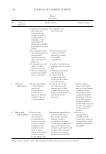
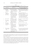
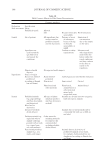
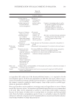
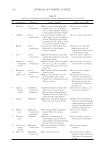
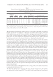
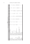
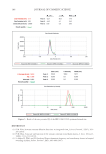
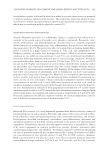
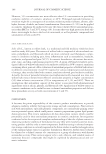
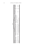
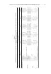
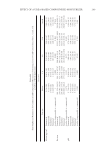
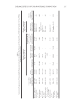
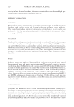
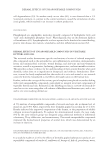
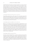
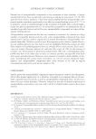
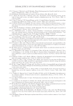
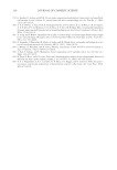
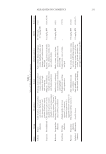
JOURNAL OF COSMETIC SCIENCE 166 4. Th e applicator was soaked in 70% medical spirit for 30 s, drained, and then impressed on the surface of medium No. 4. 5. Th e applicator was soaked in 70% medical spirit for 3 min, drained, and then impressed on the surface of medium No. 5. 6. Th e applicator was reused to apply face makeup. 7. Th e applicator was washed with soapy water, soaked in a solution for surface and tool disinfection for 30 s, drained, and then impressed on the surface of medium No. 6. 8. Th e applicator was soaked in solution for surface and tool disinfection for 3 m, drained, and then impressed on the surface of medium No. 7. 9. Th e Petri dishes were sealed, placed in an incubator, and incubated at 37°C for 24 h. 10. Af ter 24 h, the Petri dishes were removed from the incubator and subjected to photographic documentation. 11. Co lonies of microorganisms were counted using ImageJ computer software. RESULT S After using the soap, which has triclosan with antibacterial activity, the number of microorganisms present clearly decreased, but the soap did not eliminate them com- pletely in all the analyzed cases. The effectiveness of the medical spirit, the active com- pound of which is ethyl alcohol, turned out to be 100%. No microbial colonies were noted in the control sample. It can be observed, however, that the number of microbial colonies that appeared on the surface of the Petri dishes was affected by whether the applicator under examination was used to apply a dry or wet product. In the fi rst case, a natural make-up brush, which was used to apply a dry product, was analyzed. When compared with the brush with synthetic bristles and the make-up sponge that were used to apply wet products, the number of colonies in the Petri dishes was lower after impressing the dirty applicator. There were 1,162 colonies found on the surface of the Petri dish that was used for the analysis of the synthetic brush, 617 colonies of microorganisms on the surface of the Petri dish on which the sponge tested was much smaller, and 418 colonies on the Petri dish on which the natural make-up brush was tested. The an alysis of the media is presented in Table 1. Table I Analysis of the Columbia Agar Plate Culture Plate number Natural make-up brush Synthetic make-up brush Make-up sponge #1: control test No colonies (Figure 2A) No colonies (Figure 3A) No colonies (Figure 4A) #2: water 418 colonies (Figure 2A) 1,162 colonies (Figure 3A) 617 colonies (Figure 4A) #3: soap 83 colonies (Figure 2A) Nine colonies (Figure 3A) Three colonies (Figure 4A) #4: ethanol, 30 s No colonies (Figure 2B) No colonies (Figure 3B) No colonies (Figure 4B) #5: ethanol, 3 min No colonies (Figure 3B) No colonies (Figure 3B) No colonies (Figure 4B) #6: re-disinfection after using, 30 s No colonies (Figure 2B) No colonies (Figure 3B) No colonies (Figure 4B) #7: re-disinfection after using, 3 min No colonies (Figure 2B) No colonies (Figure 3B) No colonies (Figure 4B)
Purchased for the exclusive use of nofirst nolast (unknown)
From: SCC Media Library & Resource Center (library.scconline.org)