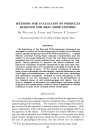
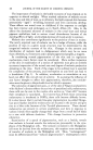
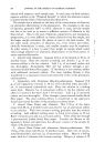
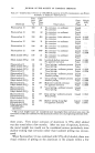
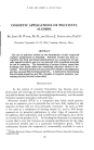
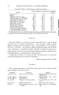
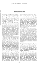
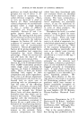
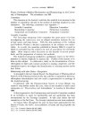
SOME ASPECTS OF MELANIN PIGMENTATION 299 . o ! Figure 1.--The epidermal melanin unit, from T. B. Fitzpatrick and A.D. Breathnach (3). N -- nucleus, M = mitrochondrion, PMS = pre-melanosome, MS -- melanosome, MG = melanin granule, G = Golgi membranes, E = endoplasmic reticulum and B.M. -- basement membrane. The development and transfer of pigmented melanin granules is illustrated in a series of five steps: I. Synthesis and condensation of the enzymic protein molecules. II. Arrangement of the molecules in structural form, leading to the development of an or- ganelle--the pre-melanosome. III. Biosynthesis of melanin and its accumulation within the pre-melanosome, proceeding through the stage of the melanosome to that of the fully synthesized melanin granule. IV. Transfer of melanin granules from melanocyte to Malpighian cells. V. Distribution of granules throughout successive epidermal levels and their final elim- ination at the surface as the result of progressive upward movement of cells from the basal layer to the stratum corneum. to the formation of melanin in the melanocyte, while melanin pigmen- tation describes not only formation of melanin but its distribution through- out the epidermal cells (Fig. 1). The dynamic process of melanin pigmentation involves two dissimilar cells (melanocyte and Malpighian cell) which appear to operate closely together as a single functioning unit each has an essential part to perform, and their roles are complementary. These two types of cells could be, considered to compose a structural and functional unit, an epidermal melanin unit (Fig. 1), analogous in some respects to other functional units such as the nephron. The epidermal melanin unit can be loosely defined as a melanocyte with an associated pool of Malpighian cells, the

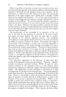
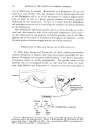
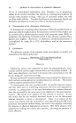
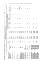
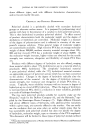
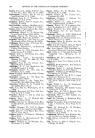
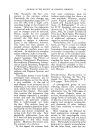
300 JOURNAL OF THE SOCIETY OF COSMETIC CHEMISTS size of which may be variable. Breathnach and Fitzpatrick (3) are not anxious to coin another term, but emphasis must be placed upon the role of the Malpighian cell as an active participant in melanin pigmentation and not upon its role as a purely passive recipient of melanin granules elaborated by the melanocyte. In fact, it is likely that the Malpighian cell may play an active role in controlling the synthesis of melanin granules by the melanocyte. After defining the epidermal melanin unit in so far as possible as a struc- tural unit, the respective roles of the individual components of this unit- (a) the melanocyte as the producer of melanin granules, and (b) the Mal- pighian cell as the vehicle of distribution throughout the epidermis----in the dynamic process of melanin pigmentation can now be examined. PRODUCTION OF MELANIN GRANULES IN MELANOCYTES In 194:9, when Lerner and Fitzpatrick (4:) were studying mammalian melanin formation, it became clear that the enzyme responsible for pro- duction of melanin was attached to particulates it was known that plant tyrosinases existed as soluble polypeptides. The specific nature of the particles was not investigated further at that time but about ten years later, Seiji, Bitbeck and Fitzpatrick (5) worked out the type and origin of • ,•o e Internal structure and TYROSINAS[ / TYROSIN [ • H ß { INTERMEDIATE COOH C•H Figure 2.--Melanogenesis in human skin, as seen in the light microscope, the electron microscope and at the molecular level. (From T. B. Fitzpatrick, M. Seiji and A.D. Mc- Gugan, New. Engl. J. Med., 265, 330, 1961). M = mitochondrion, PMS = pre-melanosome, MS = melanosome and MG = melanin granule.
Purchased for the exclusive use of nofirst nolast (unknown)
From: SCC Media Library & Resource Center (library.scconline.org)