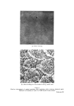
414 JOURNAL OF THE SOCIETY OF COSMETIC CHEMISTS degrees, from virtually nothing in the nordic races to almost jet black in certain Negro races. The degree of skin pigmentation is not related to numerical differences in the melanocyte population as a heavily pigmented Negro has the same number of melanocytes per unit area from a given site of skin as a white man. These racial differences in coloration must therefore be due either to greater production of pigment by the melanocytes, or to a decreased rate of destruction, or both. The pigment is produced by specialized dendritic cells, known as melanocytes, situated in the basal layer of the epidermis. It is then passed into the adjacent epidermal cells by the dendritic processes of the melanocytes. This transfer of the melanin granules to epidermal cells has been termed "cytocrine activity" (1). The pigment tends to accumulate in the upper part of the epidermal cells above the nucleus, and thus protects this vital structure from the effects of solar radiation. In white skin there is virtually a complete absence of pigment immedi- ately above the lowest layers of epidermal cells. This sudden reduction cannot be entirely accounted for by a dilution effect due to the division of the melanin-containing epidermal cells with a consequent decrease in the individual cell pigment load. In dark skin, melanin can be readily de- tected in the upper region of the epidermis and sometimes even as high as in the keratin layer. It is thought that in fair skin the pigment becomes rapidly reduced, and this results in a lightening of the colour of the melanin granules. The rapid phase of pigmentation of fair skin following exposure to sunlight is thought to be due to the photodynamic reoxidation of this reduced pigment, whereas the tanning that appears two or three weeks later is considered to be due to increased melanin production effected by actual stimulation of the melanocytes. In man both the skin and the hair are pigmented, and it is probable that the melanocyte population of the hair follicles, and of the intervening epidermis, constitute a single cell population. In animals having a heavy fur covering, the hair may or may not be pigmented but the underlying epidermis is usually devoid of pigment. This is because the overlying hair affords adequate protection from solar radiation and therefore epidermal pigmentation is not required. Pigmentation of the hair does increase the protection afforded by the keratin of the hair fibres, but the main functions of hair colour in animals is camouflage and secondary sexual characteristics. The depth at which the melanin is situated in the skin is important as this causes changes in its apparent colour. Thus pigment within the

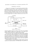
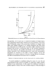
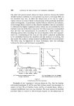
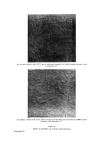
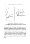
THE MELANOCYTE SYSTEM AND KERATINIZATION 415 epidermis appears as increasing shades of brown to black, whereas the same pigment deep in the dermis appears blue. The blue naevus and mon- golian spot are examples of this apparent blue colour of deeply situated melanin. The determination of natural skin colour The accurate matching, precise determination and recording of skin is difficult in dermatological practice. Photographic methods, using strictly controlled colour temperature lighting, with the emulsion developed under standardized conditions together with colour charts photographed alongside the subject, often fail to give satisfactory comparisons. A comparator method has also been used in which the skin is illuminated at the same time as a series of standard colours and the disc rotated until visual matching is obtained. Probably the best method is by the use of reflectance photometry the apparatus employed for this purpose is manufactured by Evans Electro- selenium Ltd. The principle of this technique depends on the absorption of rays of a selected wavelength directed on to the skin surface. The wave- length of the incident radiation is determined by interposing filters between the light source and the skin. The degree of absorption is estimated by measuring the light reflected back from the skin by means of a selenium cell. The current generated in the cell is recorded by a sensitive galvano- meter. The greater the quantity of the rays absorbed by the skin, the less radiation will be reflected back to the selenium cell and consequently the lower will be the galvanometer reading. For the determination of red due to blood in the skin, red light is suitable because in this region of the spectrum a good haemoglobin has a good maximum absorption. However, the maximum absorption by melanin occurs in the uv region of the spectrum and therefore the determination of the degree of brownness due to melanin was unsatisfactory when visible light was used with any of the filters supplied with the original apparatus. We therefore modified the photometer for use with uv rays. A uv light source in the form of an "overrun" projection lamp (Philips A1- 186) with a 1 mm Chance OX1 filter was substituted for the original light source to produce a radiation having a wavelength of around 13,600 21,. These rays are readily absorbed by melanin and because the selenium cell is sensitive to this range of uv light this gave a much more sensitive method of measuring the brown pigmentation of the skin (2).
Purchased for the exclusive use of nofirst nolast (unknown)
From: SCC Media Library & Resource Center (library.scconline.org)