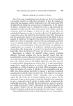
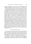
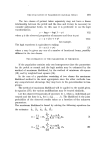
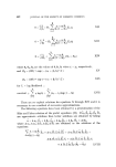
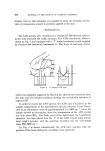
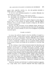
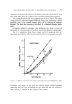
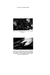
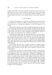
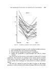
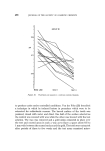
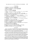
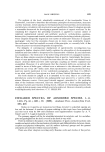
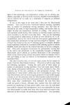
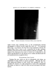
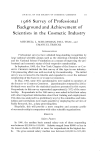
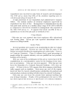
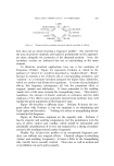
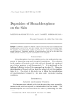
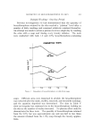
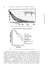
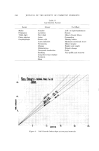
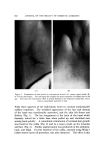
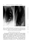
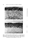
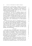
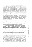
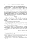
EFFECT OF TOPICAL HORMONES ON SKIN 553 Figure 3. Unilateral beard growth by topical testosterone. The left side of the face of this IM-year-old white woman was treated with the hormone for 1•/• years, while its eream vehicle was similarly applied to the right side. The mustaehe was present beforehand, as were a few wisps of hair on the chin. This again shows the purely local action of the topically applied steroid both to earlier recruitment and an increased density of functioning glands (Fig. 4). Progesterone-treated sites demonstrated effects qualitatively similar to those of testosterone but of a lower order of magnitude. Pregneno- lone acetate, while also working in the same direction, was considerably weaker in effect. Estrogens produced no observable improvement in the aged skin and, if anything, tended to suppress both hair growth and eccrine sweating in the axilla. The corticosteroid hormones unequivocally accentuated the defects of senescence. The skin became thinner, atrophic, and even more lax. Spontaneous petechiae developed in the axilla, while the forearm dis- played the identical likeness to senile purpura (Fig. 5). Hair growth in the axilla diminished. Triamcinolone acetonide suppressed eccrine function, and fluocinolone acetonide stimulated sweating. Thus, in regard to gross structure and function, testosterone, pro- gesterone, and pregnenolone, in that order, have a general rejuvenating effect on senescent skin. On the other hand, estrogen mildly accentu- ated some of the aging characteristics, while the corticosteroids pro- foundly increased the degradative alterations.

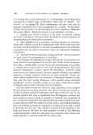
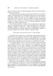
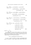
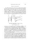
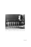
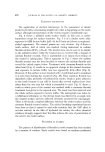
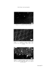
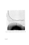
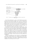
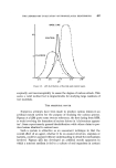
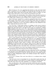
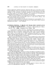
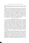
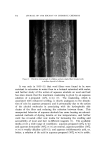
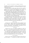
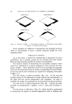
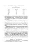
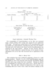
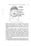
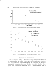
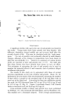
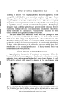
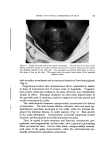
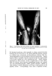
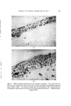
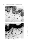
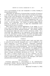
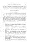
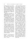
5.54 JOURNAL OF THE SOCIETY OF COSMETIC CHEMISTS .( ... (A) (B) Figure 4. The effect of topical testostcnme on eccrine function as demonstrated by use of Wada's iodine-starch-castor oil technique. Sweat droplets appear as black puneta trapped beneath the oil. The control side (A) clearly dexnonstrates the diminished activity seen in the aging axilla. (Subject is a 75-year-old white man.) The contralateral, testosterone treated area (B), however, has greatly increased sweating HISTOLOGIC CHANGES IN AGING SKIN Most impressive of the structural changes are the marked thinning and flattening of the epidermis. It is composed of cells which display great irregularity with marked discrepancy in size, shape, and staining properties. The cytoplasm of the prickle cells often condenses in the periphery, leaving perinuclear halos. The nucleus is often small and darkly stained, and the nuceoli are obscured. The orderly progression of cells from the basal layer to the surface is often disrupted, with considerable loss in cell polarity (Fig. 6A). Under the light microscopy,
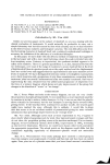
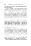
Purchased for the exclusive use of nofirst nolast (unknown)
From: SCC Media Library & Resource Center (library.scconline.org)