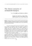
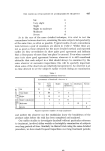
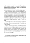
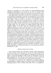
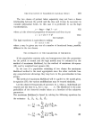
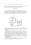
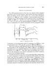
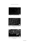
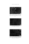
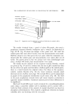
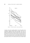
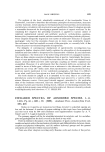
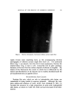
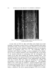
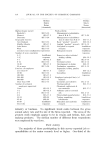
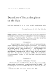
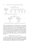
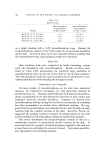
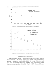
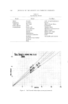
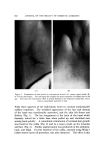
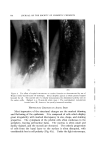
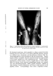
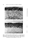
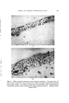
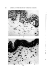
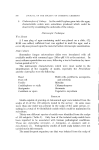
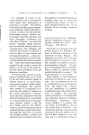
EFFECT OF TOPICAL HORMONES ON SKIX (c) Figure 8. Hormonal effect on derreal cells. The control specimen (A) exhibits the char- acteristic bipolar, spindle shaped fibroblasts, packed in parallel bundles at the upper portions of the dermis. Testosterone treatment (B) causes the cells to assrune a more blast-like, stellate form, and they are found more uniformly distributed throughout the dermis. Topical corticosteroids cause a marked diminution on the cell population (C). Either the fibrocytes are not staining or their numbers have been reduced. The relative diphosphopyridine nucleotide diaphorase (DPND) activity in the epidermis is also seen with this enzyme preparation. (Original magnification 200 X ) cytes, which are shrunken to skimpy, bipolar forms packed in parallel bundles within the Hale staining areas (Fig. 8A). HISTOLOGIC RESULTS OF STEROID APPLICATION The microscopic structural improvements following the application of hormones are far more impressive than those appredated with the unaided eye. These alterations are readily apparent in the majority of subjects (about 75%), including many individuals who showed no clinical modifications. The tenor of the testosterone effect, and that of progesterone and pregnenolone to a lesser degree, was toward a more youthful architecture. Thickening of the treated epidermis (through increase in cell size and number of cell layers) occurred to an extent which made it appear as though it had been viewed at a higher

JOURNAL OF THE SOCIETY OF COSMETIC CHEMISTS magnification than its control specimen. Regularity in cell size and shape was restored, nuclear and cytoplasmic content seemed plumper, and nucleoli became distinct. Stratification of cells throughout the epidermis was restored to the normal pattern (Fig. 6B). The basement membrane thickened perceptibly, stained more vividly, and became sharply defined and unbroken (Fig. 7B). The dermis showed increased AMP staining of deeper hue and broader distribution. There was an accompanying alteration in the morphology of the fibrocytes, which resumed a plump stellate form and were found dispersed throughout the dermis (Fig. 8B). The heavy material of basophilic degeneration appeared diluted or washed out by the increased AMP. In a similar fashion, the coarse tangles of elastic tissue appeared more separated and were pushed further away from the papillary dermis. This subepidermal zone now contained numerous fine elastic fibers which ran perpendicularly to the dermo-epidermal junction. Estrogen-treated tissues could not be distinguished from the con- trols. As with the clinical results, the corticosteroids produced degrada- tive changes easily seen by microscopic examination. The epidermis became uniformly atrophic and was often reduced to a structure of only several cell layers' depth. The basement membrane suffered further washout and became more indistinct. Dedmation of the cell popula- tion of the dermis (Fig. 8C) was accompanied by diminished AMP staining. The subepidermal orcein staining fibers were little changed following corticosteroid treatment. DISCUSSION These studies indicate that topically applied steroid hormones can produce definite alterations in aging human skin. Androgens primarily tend to ameliorate the degradative changes, while the corticosteroids further exaggerate the deterioration. Exactly how these modifications develop is not yet known, and perhaps study of the mechanisms may provide insight into the aging process itself. Cutaneous senescence at least appears to have been conveniently bracketed by this divergent action of structurally related chemicals. One fact which is clear is that the observed results are entirely local. Similar effects on distant or neighboring untreated skin do not occur. Systemic administration of the hormones in therapeutic amounts over an extended time are also ineffective (3). Of all the steroids used, only the estrogen showed significant absorption as such, with general effects
Purchased for the exclusive use of nofirst nolast (unknown)
From: SCC Media Library & Resource Center (library.scconline.org)