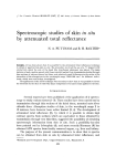
HUMAN SCALP AS HABITAT FOR MOLDS 36,5 scalp with his fingernails and by allowing some scurf to fall upon a culture medium. Each subject selected the area of scalp of his choice. It was assumed that a representative sample from all parts of the scalp was obtained. IDENTIFICATION OF MOLDS Culture Technique The culture medium used for the primary isolation was Sabouraud dextrose agar (Difco pH 5.6). To prevent the growth of yeasts and bacteria, neomycin (3 mg/ml) and nystatin (5 units/ml) were added to this medium. Primary isolations were made in Petri plates having a diameter of ,•)0 mm. Subsequently subcultures were made in smaller Petri plates (diameter 45 mm). These subcultures were grown on Sabouraud anti- biotic agar, except for the molds of the genus Penicillium, which were grown on Czapek dox agar (Difco pH 7.3). All mold subcultures were grown from ten to fourteen days in the dark at 25 øC. The cultural characteristics most useful in identification were the following: 1. Colony Growth: The rapidity of growth and the size of the colony at maturity is important. 2. Colony Surface.' The colony surface could be velvety, floccose (wooly), funiculose ,or fasciculate. Ridges and furrows oriented in a radiating or concentric manner demarcated the colony into well-defined zones (zonate). 3. Colony l•argin.' The colony margin could be undulating or entire. 4. Colony Color: The chromogenicity of the aerial parts, including hyphae, conidiophores, and conidia, was observed. Sometimes the medium surrounding the colony became colored by soluble pigments, which was an important consideration. 5. Spores: The degree of sporulation and the spore color were important in designating species. (•. t•xudates.' Droplets of liquid appearing on the surface of the colony were often seen. These droplets varied in number, clarity, and pigmentation. 7. Odor: The odors produced by molds varied considerably and were characteristic.

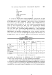
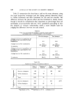
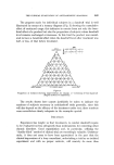
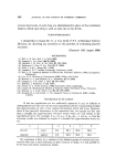
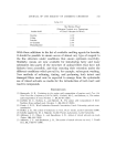
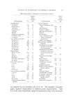
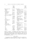
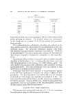
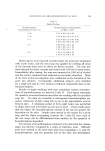
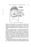
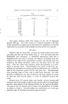
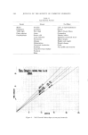
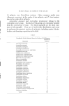
566 JOURNAL OF THE SOCIETY OF COSMETIC CHEMISTS , Undersurface of Colony: As the mold hyphae grow into the agar, characteristic colors were sometimes produced which could be observed by examining the underside of the colony. Microscopic Technique Wet Mount A 3 mm plug of agar-eontaining mold was placed on a slide, $% KOH was added, sufficient heat was applied to melt the agar, and a cover slip was pressed upon the material before microscopic examination. Slide Culture Shoemaker fungus microculture slides were inoculated with all available molds with cornmeal agar (Difco pH 6.0) as the nutrient. In most cultures sporulation was seen, follow ing a ten to fourteen day incu- bation period at 25 øC. The microscopic characteristics which were most useful in the identification of the majority of molds, especially the Penicillium and the Aspergillus •-ere the following: Head Foot-cell Vesicle Conidiophore or stalk Steriõmata Conidium or spore Hulle cells, perithecia, ascospores, and sclerotia Metulae Chlamydospores Stromata Vegetative mycdium RESULTS Molds capable of growing on Sabouraud agar were isolated from the scalps of 55 of the 100 subjects tested in the survey. In some cases more than one mold was present in the scalp of the same person, re- sulting in a total of 90 identifiable molds which could be maintained in subculture. Thirty-one different species of molds were identified from the scalps of 100 subjects (Table I). Only four of the isolated scalp molds have been reported to be associated with human pathological conditions. These are Aspergillus versicolor, A. fumigatus, A. awamori, and A. r•iyckoensis (5). Pathogenicity studies of mold scalp isolates were not carried out in this research. The most frequent organism, one that was isolated from the scalp of
Purchased for the exclusive use of nofirst nolast (unknown)
From: SCC Media Library & Resource Center (library.scconline.org)