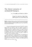
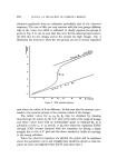
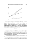
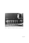
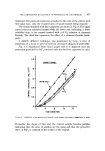
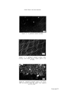
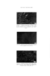
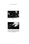
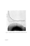
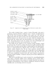
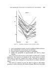
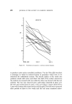
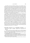
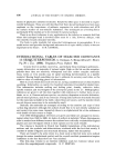
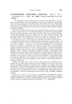
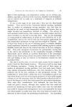
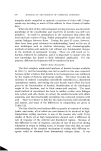
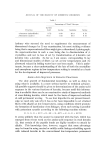
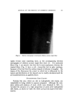
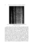
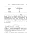
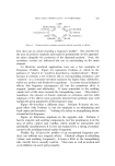
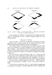
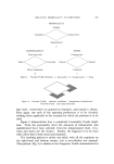
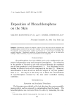
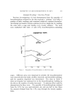
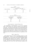
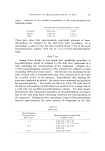
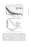
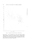
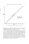
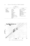
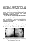
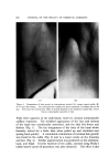
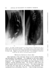
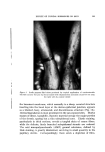
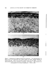
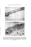
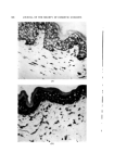
478 JOURNAL OF THE SOCIETY OF COSMETIC CHEMISTS ELECTRON MICROSCOPY The application of electron microscopy to the assessment of dental products provides a fascinating insight into what is happening on the tooth surface although interpretation of the results requires considerable care. Fig. 4 shows a polished tooth surface which, in this case, is rather featureless except for surface scratches. Fig. $ is of a similar tooth after exposure to 0.2M lactate buffer (pH--4) for 15 min and shows a roughened, etched appearance. The enamel prisms are very obvious. Fig. 6 shows a tooth surface, half of which was masked during immersion in sodium fluoride solution (0.2%: pH =4). The masked area can be seen to be similar to the polished surface while the treated area is covered with a deposit of calcium fluoride crystals. This is undesirable as it means that erosion of the enamel is taking place. This is apparent in Fig. 7 where the sodium fluoride treated area has been brushed to remove the calcium fluoride and reveal the etched region below. Treatment with stannous fluoride on the other hand (Fig. 8) results in no apparent change in the enamel structure and exposure to lactate buffer now has apparently little effect (Fig. 9). However, if this surface is now brushed with a toothbrush and re-examined it is seen that etching has occurred (Fig. 10). Thus stannous fluoride has deposited a film, probably of hydrated tin oxides, which is quite adherent to the tooth because it can still be found after several replicas have been taken from the one area but which is permeable to acid. Fig. 11 shows a tooth in which part of the enamel was masked while a stannous fluoride treatment was given to the exposed area. The mask was then removed and the whole surface exposed to lactate buffer. The micrograph was taken by a reflectance technique, i.e. the elctron beam was directed at an angle onto the enamel surface and the reflected electrons produced the micrograph. There is obviously a marked difference between the etched surface and the stannous fluoride treated surface. The critical brushing experiment has not been done so that it cannot be said with certainty that the 'cliff' effect is not due entirely to the film of hydrated tin oxides the other chemical evidence strongly suggests that this film reduces the rate of penetration of the acid to the tooth surface. FLUORIDE IN ENAMEL The pattern of distribution of fluoride in tooth enamel has been studied by a number of workers. Early results were based on analyses performed

ELECTRON MICROGRAPHS Figure 4. A polished enamel surface. Figure 5. A polished enamel surface after etching •vith 0.2M lactate buffer (pH 4) for 15 min. ' Figure •. Polished enamel surface: the lower half was maaked while the upper was exposed to 0.2 % NaF. Facing page 478
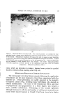
Purchased for the exclusive use of nofirst nolast (unknown)
From: SCC Media Library & Resource Center (library.scconline.org)