UV-BESTRAHLUNG DER HUMANHAUT 823 LITERATUR (1) •enilek, A., Kril, J. A., The occurrence ofurocanic acid in human sweat. Biochim. Biophys. Acta 12, 479 (1953). (2) Hall, D. A., Histidine •x-deaminase and the production of urocanic acid in the mammal. Blochem. jr. 51, 499 (1952). (3) Zannoni, V. C., La Du, B. N., Determination of histidine alphadeaminase in human stratum corneum and its absence in histidinaemia. Biochem. J. 88, 160 (1963). (4) Schwarz, E., Abbau yon Histidin zu Urocaninsiiure in der Epidermis. Untersuchungen mir L-Histidin-2 (Ring) - 14Co Blochem. Z. 334, 415 (1961). (5) •enftek, A., Kril, J. A., Hais, I. M., "Sun-screening" effect of urocanic acid. Biochim. Biophys. Acta 18, 589 (1955). (6) Krfil, J. A., Kfitovi, M., •eniiek, A., Hais, I. M., Acide urocanique dans la sueur et la r6activit• de la peau • Firradiation. Cosmgtologie 1 (2), 42 (1957). (7) Spier, H. W., Pascher, G., Analytische und physiologische Untersuchungen fiber die wasser- 1/Sslichen Inhaltsstoffe der peripheren Hornschicht (Hautoberfiiiche). Acta DermatoveneroL (•Stockholm) 2, 14 (1957). (8) Zeniiek, A., Der "sun-screening"-Effekt der Urocaninsiiure, eines neuentdeckten Bestand- teiles des menschlichen SchweiSes. Parfilm., Kosmetik 37, 350 (1956) •enfiek, A., Hais, I. M., Krfil, J. A., L'acide urocanique, antisolaire physiologique. Parrum. Costagr., Savons 9, 65 (1966). (9) Fe}tek, O., •enftek, A., Hais, I. M., Die Schutzwirkung der Urocaninsiiure im biologischen Vetsuch. Parfi#n. Kosmetik 50, 223 (1969). Baden, H. P., Pathak, M. A., The metabolism and function of urocanic acid in skin. jr. Invest. DermatoL 48, 11 (1967). Kr•l, J. A., •en•iek, A., Hais, I. M., Sweat and exercise. J. Sports Med. & Phys. Fitness 3, 105 (1963). Kril, J. A., •enfiek, A., gtrych, A., Hals, I. M., Petr{•f•ovl, O., Kalouskov{•, A., Hovorka, J., Urocaninsiiuregehalt der Epidermis bei Afrikanem und Europ•iern. Parfan. Kosmetik 48, 193 (1967). Anglin, J. H., Jones, D. H., Beret, A. I., Everett, M. A., The effect of ultraviolet light and thiol compounds on guinea pig skin histidase. J. Invest. DeramtoL 46, 34 (1966). Reaven, E. P., Cox. A. J., Histidine and Keratinization. Ibid. 45, 422 (1965). •enfiek, A., Kril,J. A., Hals, I. M., gtrych, A., Adaptive Sonnenschutzmechanismen im Nach- trag zur Sonnenbriiunung. ]. Soc. Cosmetic Chemists 20, 315 (1969). •eni.•ek, A.. Hals, I. M., •trych, A., Kril,J.A., L'antisolaire naturel est-il influenc• par Firradiation solaire ? Parfum. CostaR. Savons, 12, 382 (1969). Kril, J. A., :•enfiek, A., gtrych, Hais, Tloutfka poko3.ky a koncentrace kyseliny urokinov6 na posterolaterilni (pigmentovan•jif)a anteromediiln• (m•n• pigmentoran6) stran• pa3.e. • as. Lgk. ges., 107, 302 (1968). Hais, i. M., gtryc14, A., gpa&k, J., •en/iek, A., Kril, J. A., The increase of epidermal imida- zolacrylic acid following insolation. A study based on a group of 15 human subjects. J. Invest. DermatoL 55, 39 (1970). Kiistala, U., Mustakallio, K. K., In-vivo separation of epidermis by production of suction blisters. Lancet I, 1444 (1964). Tabor, H., Mehler, A. H., Histidase and urocanase. A. Histidase-Methods in Enzymology (N. O. Kaplan and S. P. Colowick, ed.) Vol. II, p. 231. New York, Academic Press, 1955. Baden, H. P., Pathak, M. A., Biochim. Biophys. Acta 104, 200-204 (1965). Baden, H. P., Pathak, M. A., Butler, D., Nature 210, 732-733 (1966). Merritt, A.D., Rucknagel, D. T., Silverman, M., Gatdiner, R. C.,J. Clin. Invest. 41, 1472-1483 (1962). (10) (11) (12) (13) (14) (15) (16) (17) 08) (19) (20) (21) (22) (23)

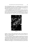
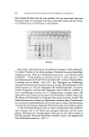
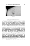
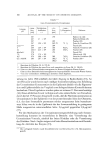
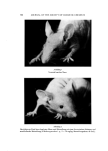
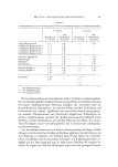
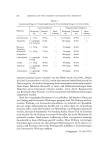
j. Soc. Cosmetic Chemists 21, 825-833 (1970) Prtifung von Kosmetik-Grundstoffen auf fototoxis&e Wirkung CHRISTIAN GLO XHUBER* Vorgetragen am 9. Mai 1970 in Ggttingen Synopsis--Phototoxicity Testing of Cosmetic Materials. Sunlight can cause chronic skin damage as well as malignancies. Certain individuals show greater sensitivity to sunlight than others (porphyria). Exogenous light damage also includes photo-allergic (Refs. 7, 21, 22) and phototoxic reactions (Refs. 1, 11). A particularly well-known phototoxic reaction is the so-called Bexloque dermatitis which is caused by furocumarins (Refs. 4,15, 20). Other phototoxic reactions have been associated with the use of tetracyclines• tars• and griseofulvin (Refs. 13, 14, 18). Animal testing procedures for assessing phototoxic activity have been used in the past (Refs. 8, 5). The method used by the author is simple. Individual hairless mice are irradiated with an Osram Ultravitalux lamp in wire cages at a distance of 50 cm. The intensity of the irradiation is attenuated through a glass plate, and it is necessary to irradiate continuously for 48 hours in order to initiate erythema in the experimental animals. The application of the test substance varies: Undiluted materials or dilute solutions are brushed on the skin of the back in other instances, the materials are injected intraperitoneally. One group of test animals is treated with the material and irradiated. A second group remains untreated but is exposed to the U.V. light. A second control group is treated with the test sub- stance but is not irradiated. After completion of the irradiation, the initial results are read, and delayed reactions are observed on the next day. The results of the tests are shown in Tables 1 and 2, which are concerned respectively with essential oils and a variety of drugs. Figure •_ shows the appearance of a normal hairless mouse, whereas Fig. 2 shows edema on the head ooe a hairless mouse after treatment with a phototoxic material and sub- sequent irradiation. Key to Table I: Horizontal: 1st Experiment 2nd Experiment Irradiation No Irradiation Irradiation No Irradiation 3 x 24 hrs. Control 4 x 24 hrs. Control Footnotes: * Two replications ** Fo•,r replications *** For light-protective agents o. B = no effect * Toxikoloõische Laboratorien der Henkel & Cie. GmbH, 4 Diisseldorf. 825
Purchased for the exclusive use of nofirst nolast (unknown)
From: SCC Media Library & Resource Center (library.scconline.org)