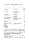
J. Soc. Cosmet. Chem., 26, 83-91 (February 1975) Aerobic Microflora of the Outer Eye Area of Women of Los Angeles, Calif. JOHN F. McCONVILLE, B.S., and DAVID W. ANDERSON, Jr., Ph.D.* Synopsis-Several reports have enumerated the MICROFLORA of used eye cosmetics. However, there is a paucity of literature describing the microflora of the OUTER EYE AREA. Understanding of the microflora of the outer skin around the eye should be useful in the development of PRESERVATIVE systems for eye cosmetics. The purpose of this paper is to contribute to the knowledge of the microflora of the outer eye as determined in selected subiects residing in Los Angeles, Calif. INTRODUCTION Knowledge of the microbiological flora present on the human face, in par- ticular the eye area, is of importance to the cosmetic industry (1-5). The formulator of products for application on the human face should consider the types and numbers of microorganisms apt to be introduced into a product by applicators and/or fingers. Awareness of the resident and transient micro- organisms found about the eye is useful in the development of ocular cos- metics in order to adequately preserve them against the survival of these microorganisms when introduced into the product by the consumer. The purpose of this work was to define the numbers and types of aerobic microorganisms normally found on the outer eye and to ascertain if poten- tially pathogenic aerobic bacteria are found as residents or transients. MATERIALS AND METHODS Two separate studies were performed on two different groups of subjects selected from laboratory personnel. Various techniques have been employed in previous studies of the microflora present on human skin. However, be- cause of the delicacy of the outer eye area, the method for sampling the micro- flora could not irritate the eve area of volunteer subjects. Also, we wanted the sampling method to simulate the application of eye area cosmetics. Thus, the •'Max Factor & Co., 1655 N. McCadden Place, Hollywood, Calif. 90028. 83

84 JOURNAL OF THE SOCIETY OF COSMETIC CHEMISTS standardized swabbing technique, although not totally consistent and re- producible, was the method of choice for sampling the microflora of the outer eye. The first study involved a single swabbing of the eyebrow, the upper eyelid margin, and the lower eyelid margin of 21 subjects, both male and female. The second study involved four consecutive swabbings of the eyebrow and upper eyelid margin of five randomly selected female subjects to determine week-to-week variations of the outer eye microflora. In all cases, the swab- bings were done during the day and all female subjects wore their usual makb- Up. The swabbing technique employed presterilized, individually wrapped polyester fiber-tipped applicators* moistened with a sterile solution of 0.5% r'o15 sorbate 80' in normal saline. The premoistened swabs were streaked four times over the area being sampled with concomitant twirling to insure full surface contact. Immediately after sampling, each swab tip was broken off into a 20 x 125-mm screw cap test tube containing 10 ml of Letheen broth ( Difco ). All sample tubes were mixed by vortex for 1 min and i ml of each sample broth was serially diluted in sterile Polysorbate 80-saline. One milliliter of each sample dilution was plated in duplicate into Trypticase Soy Agar (TSA) for bacteria and Sabouraud's Dextrose Agar (SDA) for yeast and fun- gi. TSA plates were incubated at 35øC for 48 hours and SDA plates were incubated at 28øC for 5 days. The colonies found on these plates were re- ported as count per swab for the particular area examined. Swab-broth tubes were incubated at 35øC for 24 hours and then one loop- ful of each was streaked onto a TSA plate for isolation of the bacteria present. The plates were incubated first at 35øC for 24 hours and then at room tem- perature for an additional 48 hours. Representative dissimilar colonies were gram-stained. Colonies of gram-positive spore-forming rods were con- sidered as Bacillus sp. Colonies of gram-positive nonsporeforming rods re- sembling corynebacteria were considered lipophilic diphtheroids if the TSA colony was small and translucent, and as nonlipophilic diphtheroids if the TSA colony was large and dirty white. Colonies of gram-negative rods were transferred to Eosin Methylene Blue Agar (BBL), MacConkey's Agar (BBL), Brilliant Green Agar (BBL), and Pseudomonas Isolation Agar (Difco). Isolated colonies on these differential agars were transferred to Triple Sugar Iron Agar (BBL) and Simmons Ci- trate Agar (BBL). Fermentative gram-negative rods were identified by the Pathotec "Rapid I-D System"©$ and nonfermentative gram-negative rods were identified by a scheme of characteristics according to Pickett (6). *Falcon Plastics, Los Angeles, Calif. 90045. •Atlas Chemical, Division ICI America, Wilmington, Del. 19899. $General Diagnostics Division, Warner-Lambert Company, Morris Plains, N.J. 07950.
Purchased for the exclusive use of nofirst nolast (unknown)
From: SCC Media Library & Resource Center (library.scconline.org)