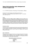
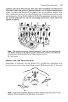
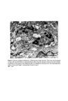
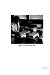
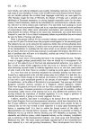
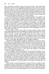
Figure 6. Human malignant melanocyte. Ultrastructural dopa reaction. The tissue was incubated in dopa after partial fixation in glutaraldehyde. Dense reaction product indicates the inter-cellular localization of tyrosinase in the Golgi saccules (*), endoplasmic reticulum near the Golgi apparatus (open arrows) and Stage I melanosomes (broad arrows). Bar: 1

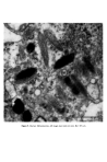
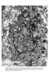
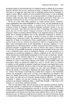
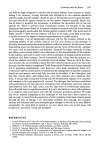
Figure 7. Melanosomes within Caucasoid keratinocyte. Basal layer keratinocyte (K) contains many complexed melanosomes (arrows) and a few single ones (open arrows). BL: basal lamina d' dendritic process of melanocyte Bar: 1
Purchased for the exclusive use of nofirst nolast (unknown)
From: SCC Media Library & Resource Center (library.scconline.org)