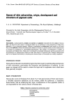
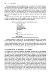
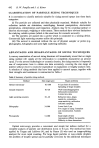
J. Soc. Cosmet. Chem. 28 621-627 (1977)¸ 1977 Society of Cosmetic Chemists of Great Britain Iauses of skin 0olouration, origin, development and struoture of pigment oells J. A. A. HUNTER Department of Dermatology, The Royal Infirmary, Edinburgh Presented at the Joint Symposium with the Pharmaceutical Society of Great Britain, "Cosmetic and Pharmacological Aspects of Colour" 9-11 November 1976, Stratford upon Avon Synopsis Haemoglobin, oxyhaemoglobin, melanin and carotene are pigments responsible for the colour of human skin. An abnormal skin colour is produced either by an imbalance in the proportion of these four pigments or by an abnormal pigment. Melanin is synthesised in melanoeytes found usually in the basal layer of the epidermis. Within the melanocytes melanin is bound to a protein matrix and the melanosomes so formed are transferred to surrounding keratinocytes. The number of melanocytes is similar in Caucasoid and Negroid skin. Black skin is produced by increased melanocytic activity associated with the production of melanosomes which are larger than those in Caucasold skin. Negroid melanosomes tend to be disposed individually in keratinocytes whereas those in Caucasoids are usually complexed. INTRODUCTION During the last decade considerable progress has been made in understanding mechanisms of skin colouration, particularly the formation and distribution of melanin. In this paper an attempt will be made to review the topic and to emphasise views which have been generally accepted. SKIN PIGMENTS Human skin varies in thickness from about 3 to 5 mm and consists of three main layers: a stratified squamous epithelium on the surface, called the epidermis a connective tissue dermis, and an underlying fatty layer. Combinations of four pigments (Table I) are responsible for the various colours of human skin. Table I. Pigments concerned in normal skin colour Haemoglobin Oxyhaemoglobin Melanin Carotene 621

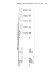
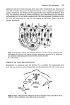
622 J. A. A. Hunter The pink appearance of untanned Caucasoid skin is due to the reddish pigment haemoglobin in the small blood vessels of the superficial dermis. If a sun tan is acquired this pink colour is partly obscured by a brown pigment, melanin, in the overlying epidermis. Melanin is, of course, also responsible for the various shades of brown seen in Negroids. Other hues are produced by the combination of these pigments with the fourth one, carotene, a yellow substance found in the subcutaneous fat and epidermis. There is no blue pigment, and when this colour is seen, it is the result of an optical effect (1). Abnormal skin colour may either be produced by an imbalance in the proportion of these four pigments, as seen for example, in cyanosis, diffuse melanosis and caro- tenaemia, or may be caused by an abnormal pigment (Table II). Table H. Some pigments responsible for abnormal skin colour HaemoglobinSproducts Methaemoglobin Sulphaemoglobin Carboxyhaemoglobin Billrubin and Biliverdin Haemosiderin Metals Gold, Silver Drugs Mepacrine, Clofazimine, Chlorpromazine Tattoo Pigments Although melanin is therefore only one of many pigments, the rest of this paper will be devoted solely to the study of mechanisms involved in pigmentation due to melanin. in emphasising structural aspects an attempt will be made to set the scene of melanin formation and disposal. Later papers will both amplify some of the points mentioned here only briefly and expand the general theme to include discussions on the biochemistry, control and significance of melanin pigmentation. CELLULAR SITE OF MELANIN SYNTHESIS The pioneer work of Bloch (2) indicated that certain branched cells in the epidermis now called melanocytes, contained an oxidising factor (dopa-oxidase) which catalysed the oxidation of the coloufiess dihydroxyphenylalanine (dopa) to an insoluble dense substance, melanin. Improvements in Bloch's dopa technique (3) led the Canadian Masson (4) to propose that the dopa positive melanocyte was the only melanin producing cell in the epidermis. Fitzpatrick, Becker, Lerner and Montgomery (5) modifying Bloch's method by using tyrosine instead of dopa as a substrate, demonstrated the presence of tyrosinase in melanocytes. Most workers today consider tyrosinase and dopa-oxidase to be identical the enzyme catalysing both tyrosine and dopa in the initial steps of melanin formation. Billingham (6) showed that the branches of melano- cytes, seen in the epidermal basal layer, travelled along the intercellular spaces between ordinary epidermal cells (keratinocytes), split frequently, and terminated in the form of 'caps' or 'end buttons' closely applied to the walls of keratinocytes. He felt that melanin granules were manufactured within the melanocyte and passed to the surrounding
Purchased for the exclusive use of nofirst nolast (unknown)
From: SCC Media Library & Resource Center (library.scconline.org)