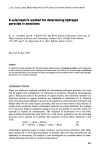
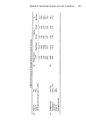
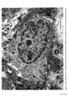
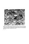
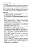
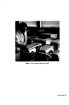
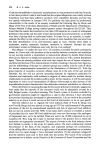
Figure 7. Melanosomes within Caucasoid keratinocyte. Basal layer keratinocyte (K) contains many complexed melanosomes (arrows) and a few single ones (open arrows). BL: basal lamina d' dendritic process of melanocyte Bar: 1

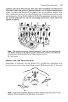
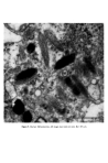
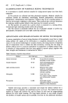
Causes of skin colouration 625 He felt that cross-linking of these filaments both at the periphery and at the centre of the melanosome was responsible for the transverse striations seen in micrographs. After formation of the protein matrix, melanin deposition gradually occurs, and the pigment accumulates on the inner membranes obscuring the characteristic periodicity of the structure. Finally the organelle becomes a uniformly dense particle without discernible internal structure. Four stages in the development of the melanosome are recognized (13): Stage I. A spherical, membrane delineated vesicle may be called a melanosome if it: (i) is shown to contain tyrosinase by electron microscopy combined with histo- chemistry or (ii) contains filaments that have a distinct periodicity of 100A. Stage II. The organelle is oval and shows numerous membranous filaments, with or without cross linking, having a distinct periodicity. Stage III. The internal structure, characteristic of Stage II has become partially obscured by electron-dense melanin. Stage IV. The oval organelle is electron-opaque without discernible internal structure in routine preparations. Stage I melanosomes are seen as spherical vesicles near the Golgi apparatus. The other stages are usually seen scattered singly throughout the cytoplasm (Fig. 5) though there is a preponderance of Stage III and Stage IV melanosomes in the dendritic pro- cesses. If preservation is good a distinct unit membrane can be seen surrounding the internal structure of the organelle (Fig. 4). Occasionally complexed melanosomes are also seen in normal melanocytes. Intracellular site of melanin synthesis Information obtained from electron microscopy, electron microscopic cytochemistry, autoradiography and cell particle fractionation supports the view that tyrosinase is synthesised on the ribsosomes. It is then transferred via the rough endoplasmic reticu- lum to the Golgi apparatus from where it is channelled via tubular elements to a focal dilatation of the smooth endoplasmic reticulum in which the coiled melanosomal matrix has independently formed. Melanisation of the structural protein can then take place and once this is completed the connection with tubular system is severed (14, 15, 16). RACIAL DIFFERENCES IN PIGMENTATION It seems extraordinary that it was not until the late nineteen sixties that Man began to understand why Negroes were black and Caucasoids white. The work of Szabo, Wolff and their colleagues (17, 18, 19) has clarified the problem and is worth summarising. There is no difference in the number of melanocytes between Negroes and Caucasoids (20). There are, however, fewer melanosomes in the melanocytes and keratinocytes of Caucasoids and Mongoloids. Of those present in the melanocyte most are in Stages I, II and III. Those in the keratinocytes are in Stage IV but tend to be grouped in membrane- limited organelles to form 'melanosome complexes'. The appearance is different in Negroids and Australian aborigines. Here there are more melanosomes in the melanocytes and keratinocytes and a high proportion of melanosomes are seen at the IVth stage of
Purchased for the exclusive use of nofirst nolast (unknown)
From: SCC Media Library & Resource Center (library.scconline.org)