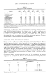
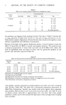
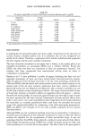
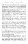
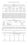
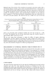
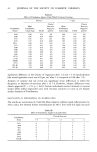
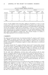
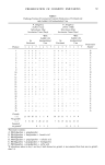
STRATUM CORNEUM FUNCTION 13 Table I Transepidermal Water Loss (gm-2hr -') at Three Sites in Four Subjects Before and After 4% Poldine Methanesulphonate Solution (95% ethanol). Means of Six Readings at 5 min-intervals with Standard Deviations Before Poldine After Poldine Subject Forearm Upper Arm Abdomen Forearm Upper Arm Abdomen 1 3.25 _+ 1.08 3.88 _+ 1.20 3.56 _+ 1.13 2.77 _+ 0.75 3.25 _+ 0.82 4.68 _+ 0.42 2 1.92 _+ 0.79 2.87 _+ 1.04 3.62 _+ 0.48 2.98 + 0.52 2.62 _+ 0.82 3.47 _+ 0.63 3 3.40 _+ 2.57 2.55 _+ 0.50 2.87 _+ 0.34 2.02 _+ 1.23 1.88 _+ 0.86 2.87 _+ 0.58 4 2.68 _+ 0.74 2.82 _+ 1.03 2.77 _+ 0.32 3.17 _+ 2.03 2.16 _+ 1.06 2.28 _+ 0.45 the same sites (forearm, upper arm, abdomen) showed moderately good reproducibil- ity (Table I). Table I also demonstrates that under these conditions the use of an antiperspirant is unnecessary, as sweating is minimal and constant. The ease and rapidity of measurements with the evaporimeter permits multiple readings on the same subject within a short period. This has enabled us to take TEWL readings in psoriatic patients attending the clinic (demonstrating enhanced water loss in the apparently uninvolved paralesional skin, Table II). The measurement of TEWL has been used as Table II Transepidermal Water Loss in Psoriatic Subjects and Controls Number of Subjects Sites Subjects TEWL (g m -2 hr ') + SD Controls Forearm 9 3.32 -+ 1.87 Psoriatics Lesions on Arms 6 10.60 _+ 6.64 Psoriatics Immediately paralesional 6 6.22 _+ 3.06 Psoriatics Uninvolved sites 6 3.78 + 2.27 an index of SC hydration (4) in normal skin in vitro. Our own experience suggests that, in vivo, with occlusive applications there is a decrease in water loss with increased hydration, though as yet we are uncertain as to the linearity of the response (Table III). It is interesting to note that other of our observations suggest that thick and compact Table III Measurement of Transepidermal Water Loss (g m-2 hr-•) after Application of White Soft Paraffin (WSP) Minutes after application of WSP Subject 0 30 120 180 1 4.5 2.5 3.5 1.0 2 3.75 3.0 2.5 1.5 3 4.5 3.5 4.0 2.5 4 4.25 3.0 2.5 2.5 5 5.25 2.5 2.5 2.5 6 2.5 2.5 2.5 2.0 Mean 4.13 2.83 2.92 2.0 S.D. 0.93 0.41 0.66 0.63

14 JOURNAL OF THE SOCIETY OF COSMETIC CHEMISTS SC is in itself not a particularly efficient water barrier, as water loss from poldine- treated palms, and from patients with ichthyosis, is increased. We have recently measured TEWL from eight patients with a variety of ichthyotic disorders. The mean and standard deviation for this group are 8.29 and 4.96 (gm -2 hr-•). The large amount of data that this technique will generate should teach us much about the nature of the barrier and perhaps lead to efficient ways in which to manipulate it. ASSESSMENT OF SC PENETRATION BY TOPICALLY APPLIED SUBSTANCES There are few techniques that permit tracing the permeation of the SC by substances applied to the skin in vivo. Admittedly, there are many ways of investigating complete percutaneous absorption, and, as the SC provides the skin with the bulk of its barrier property, measuring percutaneous absorption provides a measure of pefcomeal absorption. For some purposes, however, the ability to pinpoint the site and rates of permeation through the SC itself would be extremely useful. We described a technique which satisfies this requirement (5), but unfortunately it is expensive in both time and money and is only applicable to certain substances. It involves taking serial skin surface biopsies (6) (strips of stratum comeurn removed intact with a cyanoacrylate adhesive) at increasing depths and from adjacent sites, at different times after treatment of the sites with the substance under test. Each skin surface biopsy is then examined in a scanning electron microscope, equipped with an electron probe X-ray microanalysis facility. By determining the amount of the substance at each level within the SC at differing times, a kinetic profile of the permeation is obtained. In addition, it is possible to obtain an "X-ray map" of the area examined, which demonstrates the absorption visually, enabling the channels of absorption to be identified. The limitations of this method include the fact that the technique only provides an elemental analysis for elements above atomic number 10 and for accurate determina- tions the particular element has to be present at the site of examination in amounts in excess of 0.1%. We have traced the permeation of sulpher, zinc, and lead using this method, but have been unsuccessful in locating fluorinated corticosteroids-- presumably because the fluorine is present in too small a concentration. It seems clear that a more universally applicable and less demanding method is required, and other analytical techniques are being explored. MEASURING THE MECHANICAL BARRIER FUNCTION It is difficult to disentangle hardness from elasticity. Elasticity of the SC can be tested in vitro in the horizontal dimension under controlled environmental conditions, using some type of dynamometer (7,8). The testing of this property of elasticity does not give much information, however, about the important function of protection against mechanical traumata. An apparatus capable of measuring point penetrability of the SC for use in vivo has been constructed by our group for this purpose (9). The principle is to record the force required to move a needle from a point just above the surface of the skin, a range of distances from 5-50/,t into the SC. The needle moves extremely rapidly (4 m/sec) and needles of differing point diameters can be used. These conditions largely preclude other skin components from participation in the resistance
Purchased for the exclusive use of nofirst nolast (unknown)
From: SCC Media Library & Resource Center (library.scconline.org)