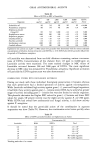
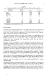
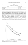
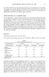
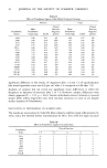
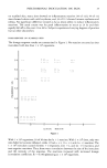
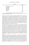
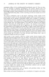
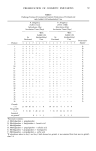
STRATUM CORNEUM FUNCTION 15 Table IV Effect of Removal of Stratum Corneum by Skin Surface Biopsy (SSB) on Penetrability of SC. 1 •t Needle Tip Used With 90-100 •t Displacement State of SC Force (mgs) + SD Before removal 32.67 + 3.06 After 4 SSBs 22.67 + 2.52 After 6 SSBs 12.67 _+ 8.02 to the needle. In order to demonstrate that it is the SC alone that accounts for the resistance to penetration in the system, we have performed two experiments in vivo. The experiments have shown that partial removal of trhe SC greatly decreases the force required for penetration, and that hydration of the SC also reduces the force required considerably (Table IV). In vitro experiments with isolated sheets of stratum corneum also show a decrease in force necessary to penetrate the SC with increasing hydration (Figure 1). It is interesting to note that this curve resembles the curves obtained by plotting electrical impedance against level of hydration in vitro and the curves obtained by plotting elastic constant against hydration level (10,11). It is interesting to note that when the force against time curve was plotted in the vivo experiments there was a dramatic change after hydration, although these changes did not accurately reproduce those found in vitro. It should be said that it appears that both penetration 25 • 15 10 05 10 20 30 40 50 60 70 80 90 100 110 120 Figure 1. In vitro determinations of point penetrability (G) in three samples of stratum corneum obtained from thighs of normal subjects with increasing hydration.

16 JOURNAL OF THE SOCIETY OF COSMETIC CHEMISTS and indentation of the SC is involved in the responses to the movement of the needle. This, in our opinion, does not invalidate the measurement, as it should be emphasized that we regard it as a measure of mechanical protectivity, rather than a measure of a pure physical property. ASSESSING THE ANTIMICROBIAL BARRIER FUNCTION OF THE SC Examination of the antimicrobial barrier property of the SC is impractical at the time of writing. In the first place, the ethical nature of the experiments would be suspect, as to make the exercise worthwhile, pathogenic microorganisms must be used. Secondly, it is virtually impossible to examine the SC under a set of standardized conditions which would minimize the contribution of sweat or sebum to the antibacterial barrier function of the SC. We are in the process of evaluating a simple in vitro test for assessing this important function of the SC, in which samples of intact SC, removed by skin surface biopsy, are treated with pure cultures of either pathogenic staphylococci or streptococci or dermatophyte fungi. After incubation for 2-14 days, the skin surface biopsies are examined microscopically, either by staining (with PAS or Grams stain), or by scanning electron microscopy. The density of the colonies, and the degree of damage caused by the invading microorganisms to the structure of the SC sheet, are indicative of the success of the particular pathogen in establishing a foothold within the horny layer. So far we have established that it is possible to use the samples of SC obtained by skin surface biopsy in this way, and that there is an intersubject variability in the degree of success for particular microorganisms. Our task now is to quantirate the observations, determine their reproducibility, and test the clinical correlation with susceptibility to infection. THE MEASUREMENT OF DESQUAMATION We have to distinguish between measurement of SC renewal time and measurement of rate of desquamation. The rate of cell loss from the surface may vary from day to day, and this variability may not be evident in determinations of renewal time. The renewal time may be accurately determined by use of a fluorescent dye that is substantive to the stratum comeurn, such as tetrachlorsalicylanilide (12) or dansyl chloride (13). There have been few attempts at measuring the desquamation rate more directly. One method has employed a resin to remove the superficial horny cells (14), but it is difficult to obtain an accurate absolute rate from this technique. Our own experiments have contrasted a method which measures desquamation when the surface is minimally disturbed (passive desquamation) with a method which assesses the loss of corneocytes after a standardized mechanical stimulus (forced desquamation). The mechanical stimulus is provided by a hand held scrub apparatus (15) based on the machine described by Nicholls and Marks (16). An electrically driven, rotating paddle impinges on the skin surface at a standard pressure for a set period, and the detached corneocytes are liberated into a solution of 0.05% Triton © x 100 in phosphate buffer (pH 7.2). The corneocytes are then removed from the chamber surrounding the paddle and counted in a haemocytometer chamber. Passive desquamation is measured with the help of specially constructed chambers made of glass fiber walls and perspex. The chambers have an internal diameter of 2 cm and there is a silicone rubber bung in the
Purchased for the exclusive use of nofirst nolast (unknown)
From: SCC Media Library & Resource Center (library.scconline.org)