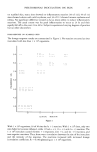
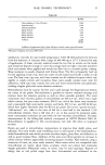
PSEUDOMONAS INOCULATION ON SKIN 21 intradermal inoculation of a 106 suspension in two subjects resulted in localized cellulitis and fever. It is well known that purulent necrotic ulcerations follow intradermal injection of Pseudomonas in animals. Again, it is hard to imagine how comparable inoculation could occur under natural circumstances. There is now, as never before, greater anxiety concerning environmental dangers, especially man-made ones. In the midst of heated controversy and vivid charges, it is important to obtain sound information which will enable dispassionate persons to form reasonable judgments concerning the hazard posed by the artifacts of civiliza- tion. Pseudomonas horror stories have been well publicized and fear of the organism is at an all time high. How serious is the situation? Cosmetic manufacturers, for example, are enjoined to ensure zero levels of Pseudomonas in their diverse products, not only those intended for use in the eye area. Further, it is urged that a product should contain sufficient preservative to ensure that it remains free of Pseudomonas during its entire use life. Are these requirements reasonable and sound? The work which we shall now report was undertaken to obtain objective data on the hazards to normal skin posed by P. aeruginosa. In addition, we studied Pseudomonas ce]gaciae, which had a reputation similar to P. aeruginosa as an opportunistic pathogen (7). SUBJECTS Healthy young adult college students ages 18-25, served as paid volunteers. Informed consent was obtained. Each study was first performed on the authors and research associates before exposing volunteers. PSEUDOMONAS STRAINS Ten strains of P. aeruginosa were obtained from the special pathogens division of the Center for Disease Control. These include six strains cultured from the skin and pool water from the aforementioned epidemics. These strains were coded 9106, 9171, 9206, 9104, 9152, and 9151. The first four are serotype 11, while 9151 and 9152 are serotype 4. The remaining strains were: serotype 11, obtained from the sputum of a hospitalized patient (strain EIA), serotype 1, (strain 9220) obtained from a water faucet, serotype 5 (strain 9190) and serotype 10, obtained by tracheal aspiration of a hospitalized patient (ff9436). In addition two strains, Duhring Laboratories strains//1 and//2, were obtained from pseudomonal interdigital athletes foot. P. aeruginosa var. hemolyticus (strain 10752) isolated from a patient with septicemia and skin lesions and three strains of P. cepaciae (strains 17759, 205608 and 27515) were obtained from the American Type Culture Collection. Prior to inoculation, all strains were subcultured every 24 hr on sheep blood agar for several passages and injected subcutaneously in rabbits to ensure virulence. Saline suspensions of fresh 24-hr subcultures were used. INOCULATION OF PSEUDOMONAS ON INTACT SKIN Forty-four male volunteers participated in this study. A 0.1-ml saline suspension containing I x 10 7 organisms/ml was applied to 4 x 3 cm areas on the volar forearm. Initially, sites were covered with impermeable plastic film (Saran ¾qrap ©) and

22 JOURNAL OF THE SOCIETY OF COSMETIC CHEMISTS overlapping strips of impermeable plastic tape. When clinical reactions failed to occur, we adopted the super-hydration technique used in our previous study. Pads of non-woven cotton cloth (Webril ©, Curity) soaked in sterile water were applied over the inoculation site and these were then occluded under impermeable plastic film and tape. Each strain was applied to two or three sites on each forearm with an uninoculated control site on each arm. One side was cultured after 48 hr and the other after 7 d. Clinical reactions were graded as follows: 0 = normal, intact skin except for soggy white moisture, response similar to control sites 1 = less than 10 papules or pustules 2 = 10-20 papules or pustules with mild surrounding erythema 3 = more than 20 papules or pustules and intense erythema 4 = confluent pustules and intense erythema 5 = erosion and ulceration INOCULATION OF PSEUDOMONAS ON SCARIFIED SKIN The purpose of these studies was to assess the virulence of various strains on skin in which the horny layer barrier was artifically breached. Superficial scarifications were made with a thirty-gauge needle according to the method of Frosch & Kligman (four parallel scratches in one direction and another four at right angles). The scratches were just deep enough to cleave the epidermis and cause pin-point bleeding from capillaries in the papillary dermis. Eight scarifications in two rows of four each could be easily made on each forearm. In the first experiment, eight strains of Pseuclomonas were tested on seven subjects (EIA, 9106, 9152, 9171, and 9436 from the Center for Disease Control, 10752 and 27515 from the American Type Culture Center, and Duhring Laboratories strain//1). A 0.1-ml saline suspension (1 x 10' organisms/ml) obtained from a 24-hr subculture was inoculated into duplicate sites, gently rubbed in with a sterile teflon rod and allowed to dry. The site was immediately covered with a semi-occlusive dressing (Band-Aid ©, Johnson & Johnson). Sites were examined at 24 and 48 hr. The clinical reactions were graded on the following scale: 0 = no reaction 1 = definite edema, mild erythema 2 = intense edema and intense erythema 3 = edema and erythema extending beyond the borders of the scarification 4 = edema and erythema extending beyond the borders of the scarification with pustule formation Duplicate readings at 24 and 48 hr were averaged. CULTURE TECHNIQUE The standard detergent-scrub technique of Williamson & Kligman was used. In brief, a 3.8-cm 2 area of skin enclosed by a sterile glass cylinder is scrubbed for 1 rain with
Purchased for the exclusive use of nofirst nolast (unknown)
From: SCC Media Library & Resource Center (library.scconline.org)