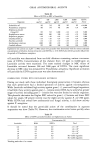
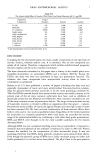
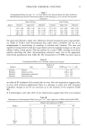
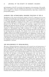
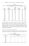
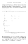
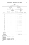
STRATUM CORNEUM FUNCTION 17 flattened tops of the devices. The chambers are attached to the skin surface with a non-irritant adhesive material, used in stoma care (Stomahesive ©) for a 2-day period, at the end of which the same buffered detergent solution as used above is injected into the chamber through the silicone rubber bung, and then withdrawn for counting the number of corneocytes present, using haemocytometer chambers. In one study we have completed (17) the forced and passive desquamation rates were determined for five sites in the same subjects. The results are set out in Table V. It can be seen that Table V Corneocyte Loss From Five Sites in Eight Normal Subjects Determined by Cl.amber (Passive) and Scrub (Forced) Techniques. Means _+ Standard Deviations. The Scrubs Were For 10 sec Chamber Scrub Sites (Corneocytes/cm2/hr) (Corneocytes/cm2/10 sec) Forearm 1309 + 328 104243 + 15570 Upper Arm 795 + 141 63660 + 5893 Abdomen 646 _+ 84.9 55623 + 8215 Thigh 878 + 140 67638 + 4736 Back 1094 + 290 81962 + 12929 there is an extremely good correlation between the two sets of data (p = 0.98, P 0.01). The absolute rate of cell loss determined using the chamber method is approximately 1000 corneocytes/cm2/hr. We are now using these techniques to assess the effects of agents enhancing desquamation and have detected interesting and subtle changes from salicylic acid preparations, which we will be reporting shortly. We believe that the two techniques should considerably help the understanding of the kinetics of cell loss from the SC and may aid in the evaluation of agents for enhancing desquamation. MEASUREMENT OF INTERNAL BINDING FORCES WITHIN THE SC The mechanism which allows the steady loss of corneocytes from the surface is as yet undiscovered. Whatever the biochemical mechanism it is accompanied by a decrease in binding force within the SC (intracorneal cohesion) (18) and measurement of this provides information about the process. In addition, we have shown that assessment of this parameter may be useful in the evaluation of hydrating agents (19). The technique for the measurement of intracorneal cohesion has been described previously (16) and depends essentially on recording the gram force required to distract from the skin surface a piston stuck to the skin with a cyanoacrylate adhesive. CONCLUSIONS The tests of function described are the ones that the Cardiff group has devised and/or evaluated and used. The most important functions are measured by them and, although other useful tests undoubtedly will be described, it is unlikely that other less important functions will be assessed. Certainly the techniques employed are capable of improvement and we are striving to that end. Skin disease alters stratum corneum

18 JOURNAL OF THE SOCIETY OF COSMETIC CHEMISTS function and this altered function may itself cause other changes within the skin. It seems intrinsically unlikely that a profile of measurements of function will give much help in the diagnostic sense--although they may do so occasionally. But it is likely that such a profile will aid in the management of patients by allowing accurate and quantifiable assessment of their progress, by determining the likelihood of serious secondary events from the altered barrier and by permitting treatment designed to rectify the barrier fault. REFERENCES (1) H. Baker and A.M. Kligman, Measurement of transepidermal water loss by electrical hygrometry, Arch. Dermatol., 96, 441-452 (1967). (2) F. R. Bettley and K. A. Grice, A method for measuring the transepidermal water loss and a means of inactivating sweat glands, Br.J. DermatoL, 77,627-638 (1965). (3) G. E. Nilsson, Measurement of water exchange through skin, Med. Bio. Eng. ½omput., 15, no. 3, 209-218 (1977). (4) A.J. Quattrone and K. Laden, Physical techniques for assessing skin moisturization,.J. $oc. Cosmet. Chem., 27, 607-623 (1976). (5) C. S. King, N. Moore, R. Marks and S. Nicholls, Preliminary studies into percorneal penetration and elemental content of the stratum corneum using X-ray microanalysis, Arch. Dermatol. Res., 263, 251-265 (1978). (6) R. Marks and R. P. R. Dawber, Skin surface biopsy, an improved technique for examination of the horny layer, Br.J. Dermatol., 84, 117-123 (1971). (7) P. Agache, J. P. Boyer and R. Laurent, Biochemical properties and microscopic morphology of human stratum corneum incubated in a wet pad in vitro, Arch. Dermatol. Res., 246, 271-283 (1973). (8) J. D. Middleton, The effect of temperature on extensibility of isolated stratum corneum and its relation to skin chapping, Br.J. Dermatol., 81,717-721 (1969). (9) S. Nicholls, C. S. King, E. Guibarra and R. Marks, Measurement of point deformation of human skin in vivo-contribution of the stratum corneum,J. Invest. DermatoL,70, No. 4, 227 (1978). (10) B. F. van Duzee, The influence of water content, chemical treatment and temperature on the rheological properties of the stratum corneum, J. Invest. DermatoL,71, No. 2, 140-144 (1978). (11) S. D. Campbell, Hydration characteristic and electrical resistivity of the stratum corneum using a non-invasive four point microelectrode method,J. Invest. DermatoL, 69, No. 3, 290-295 (1977). (12) H. Baker and A.M. Kligman, Technique for estimating turnover time of human stratum corneum, Arch. DermatoL, 95,408-411 (1967) (13) L. H. Jansen, M. T. Hojyo-Tomoko and A.M. Kligman, Improved fluorescence staining technique for estimating turnover of the human stratum corneum, Br.J. DermatoL, 90, 9-12 (1974). (14) M. S. Christensen, S. Nacht, S. L. Kantor and E. H. Gans, A method for measuring desquamation and its use for assessing the effects of some common exfoliants, J. Invest. DermatoL, 71, No. 5, 289-294 (1978). (15) E.J. Guibarra, An instrument for the stimulated release of corneocytes, Bioengineering and the skin, 1, No. 3, 17-19 (1979). (16) S. Nicholls and R. Marks, Novel techniques for the estimation of intracorneal cohesion in vivo, Br.J. Dermatol., 96, 595-602 (1977). (17) D. Roberts and R. Marks, The determination of regional age variations in the rate of desquamation. In Press. (18) C. S. King, S. P. Barton, S. Nicholls and R. Marks, Change in properties of the stratum corneum as a function of depth, Br.J. Dermatol., 100, 165-172 (1979). (19) R. Marks, Techniques for the evaluation of emollients and keratolytics, J. Soc. Cosmet. Chem., 29, 433-440 (1978).
Purchased for the exclusive use of nofirst nolast (unknown)
From: SCC Media Library & Resource Center (library.scconline.org)