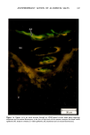
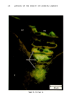
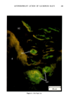
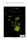
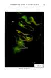
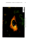
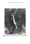
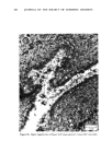
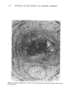
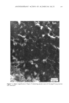
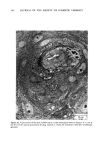
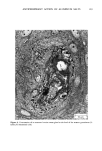
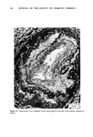
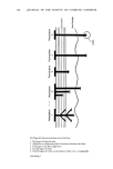
ANTIPERSPIRANT ACTION OF ALUMINUM SALTS 203 studied. The relatively superficial location of the AZAP plug nevertheless still pertains when one recognizes that the distance from the skin surface to the deepest area of fluorescence in this gland, is only ca. 70 •tm. Figure 2 is a view of a different sweat gland, showing the aluminum-and zirconium- containing plug at the most distal point of the sweat duct seated in the sweat gland ostium. Figures 3a and 3b depict yet a third gland, demonstrating the presence of aluminum and zirconlure in the intradermal duct. Three of the twelve glands studied revealed that the metals had penetrated this deeply into the sweat duct. The fluorescent area is white, and is a result of deliberate film overexposure in an attempt to elucidate otherwise dark tissue detail. Whether or not the AZAP plug in this gland had, in the living state, completely filled the lumen is conjectural, but probably unlikely, in that the morin technique is so sensitive. In any event, this would be an example of an inhibited sweat gland which was not restored to active function after Scotch tape stripping away the stratum corneum, and it indicates that the AZAP could in some instances penetrate deeply into the duct. Figure 4 is an example of an untreated sweat gland. This control tissue was histologically processed identically to the treated tissue. Fluorescence was never observed in the control sweat glands. B. OBSERVATIONS MADE USING TRANSMISSION ELECTRON MICROSCOPY The pertinent aspects of the ultrastructure of ACH-treated sweat glands in the regions where no plug material was found have been described previously (2). Those obser- vations were found to apply to AZAP-treated glands as well. The particularly noteworthy points were that the lumen of the duct was characteristically slightly dilated and essentially devoid of the products of sweat secretion, and that there was no evidence of membrane damage or cellular disruption. Figures 5-8 are transmission electron micrographs of AZAP-inhibited sweat glands. The glands were sectioned parallel to the skin surface. Figure 5a is a view of an AZAP-treated gland at the level of the viable epidermis, approximately 60/xm from the skin surface and relatively close to Lhe stratum comeurn layer as evidenced by some keratohyalin granules. For the most part, the lumen of this duct appeared empty. However, higher magnification (Figure 5b) revealed a number of small amorphous electron-dense masses which are believed to be the deepest manifestation of AZAP plug material for this gland. Additional sections of this sweat gland taken at levels progressively closer to the skin surface did not show the material to be present in amounts significantly greater than that seen in Figure 5b. Figures 6a-6c are an example of the sweat gland ostium, only 5 /xm from the skin surface. The extreme difficulty in thin-sectioning at this level so close to the skin surface invariably interfered with obtaining an anatomically well defined representa- tion of the duct opening onto the surface. Examination of Figure 6a revealed many small electron dense masses scattered throughout the area which represents the ostium. It is presumed that, in the in vivo state, these bodies were one continuous mass or plug which completely filled the pore. This presumption is based on previously described Scotch tape stripping studies which showed AZAP's site of action to be superficial, and on fluorescence micrographs such as Figures ! and 2 which also depicted the

204 JOURNAL OF THE SOCIETY OF COSMETIC CHEMISTS
Purchased for the exclusive use of nofirst nolast (unknown)
From: SCC Media Library & Resource Center (library.scconline.org)