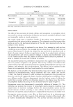
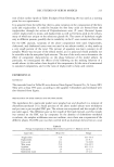
ANTI-CELLULITE ACTIVITY 203 b2 •3 Figure 1. Visualization by sonograph of the three layers of the skin (A) Skin slice by B-mode sonography with a 20-MHz probe. (B) Computed image from grabbed images and contours. 3. Mechanical characteristics of the skin: Cutometry Measurements (three measurements on each thigh) were performed with the SEM 575 © cutometer (Courage & Khazaka) on the external part of the thigh (7). The measurements were conducted in an air-conditioned room (22+/-2øC, HR 50+/-10%) after 20 min- utes of rest period. The time-strain mode was used with an elementary load cycle consisting of an instan- taneous deformation by a 500-mbar negative pressure, maintained for three seconds and followed by a three-second relaxation period, with a probe of 6-mm diameter. Three repetitions of this cycle were performed. Suction was applied with a 6-ram diameter probe. Mean Dermal Thickness: 1.86 mm Mean Dermal Echogenicity: 212.91 Dermal Texture: 31.21 Mean Hypodermal Thickness: 9.51mm Mean Hypodermal Echogenicity: 73.36 Hypodermal Texture: 27.87 Hypodermis cellulitic ratio: 7.69% Figure 2. Mean profile of computed image and quantification parameters: Delineation of the different layers allowing calculation of the different parameters.

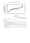
204 JOURNAL OF COSMETIC SCIENCE The studied parameters were: © Uf: maximum extensibility of the skin (or total deformation) © Ue: instant vertical extensibility (or elasticity) (Ue represents the instant deformation of the skin immediately after the application of the strain. This parameter reflects the elastic properties of the skin.) © Uv: viscoelasticity (or plasticity) extensibility (Uv represents the delayed deformation of the skin and corresponds to slower deforma- tion due to the intracutaneous movements of viscous type.) © Ur: immediate retraction (or elastic return) © Ua: total retraction © Ur/Uf: biological elasticity (Ur/Uf reflects the immediate recovering ability of the skin after the end of the strain.) © Uv/Ue: visoelasticity rate © Ua/Uf: recovery rate © R: residual deformation (R is the residual deformation observed at the end of the "off-time" period. R may be an indicator of the existence of a hysteresa phenomenon.) 4. Flowmetry of the skin perfusion: Laser Doppler flowmetry (8) Cutaneous blood flow was determined for each thigh by laser Doppler fiowmetry using the LISCA PIM II apparatus. Measurements were carried out according to the guidelines edited by the Standardization Group of the European Society of Contact Dermatitis. The measurements were conducted in an air-conditioned room (22+/-2øC, RH 50+/-10%) after 20 minutes of rest period. Skin perfusion was calculated from the image (square area of 6 cm x 6 cm) via the average value of the image's pixels. The homogeneity of the microcirculation of the skin was estimated by the standard deviation calculated from the whole laser Doppler image. ANALYSIS OF RESULTS One-way ANOVA and Student's t-test were used to determine the significance of the results the level of significance was set at p 0.05. The comparison between placebo and product was performed on the differences Tx - TO or on the means when there was no significant difference between the two thighs at TO. RESULTS MACRORELIEF OF THE SKIN With the product, the decrease of the skin macrorelief reached -53.1% at T84 days (versus -14% with the placebo). The improvement of the macrorelief was consistent throughout the study and is statistically significant at each time point. The results obtained are presented Table I.
Purchased for the exclusive use of nofirst nolast (unknown)
From: SCC Media Library & Resource Center (library.scconline.org)