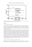
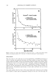
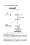
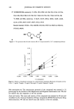
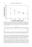
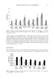
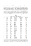
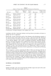
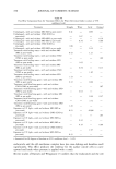
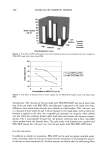
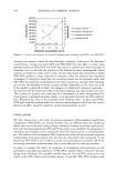
134 JOURNAL OF COSMETIC SCIENCE ering skin-whitening cosmetic preparations. In cosmetic preparations, inhibitors such as kojic acid, arbutin, ascorbic acid, and licorice extracts have been used as whitening ingredients (2). Plant extracts having an inhibitory effect on melanogenesis may be a good choice for cosmetic purposes because of their relatively few side effects. Therefore, we screened 285 plant extracts for their inhibitory activity on tyrosinase (3). Among the plant extracts, R. mori extracts showed potent inhibition activity on tyrosinase and melanin synthesis but did not inhibit tyrosinase synthesis and gene expression by zymography and RT- PCR, respectively. R. mori extracts showed inhibition of pigmentation and no toxicity in animal tests. Rlorus a/ba L. and other plants of the same genus have been used as antiphlogistics, diuretics, and expectorants in Chinese herbal medicine. R. mori (young twigs of Rlorus alba L.) is harvested in the early spring and used in folk remedies in Korea. Although the constituents of Horus alba L., such as flavonoids, coumarines, and stilbenes (4), have been studied by many investigators and isolated, few reports on the usage of cosmetic whitening ingredients have been published. In this study, we purified and identified an active compound from the R. mori extract, 2,3',4,5'-tetrahydroxystilbene(2-oxyresveratrol). A naturally occurring compound par- ticularly found in Horus alba, it showed inhibition activity on tyrosinase (IC5o = 0.23 •g/ml). Also, it inhibited tyrosinase activity in a competitive manner (Ki = 1.5 x 10 -6 M) when L-tyrosine was used as a substrate. MATERIALS AND METHODS PREPARATION OF PLANT EXTRACTS We prepared the R. mori extracts for anti-melanogenic ingredients. R. mori was extracted by a mixture of ethyl alcohol and water (EtOH:H20 = 70:30) and dried to powder. This powder was dissolved in 1,3 butylene glycol and used for this study. TYROSINASE INHIBITION For the assay, the test reaction mixture was prepared by adding 0.5 ml of R. mori extracts, to which 250 units of mushroom tyrosinase (Sigma, Saint Louis, MO) had been added, to 0.5 ml of L-tyrosine (0.1 mg/ml) or 0.5 ml of 50 mM sodium phosphate buffer (pH 6.8). After incubation for ten minutes at 37øC, we measured tyrosinase activity by absorbency at 475 nm. We determined the effect of the test sample on tyrosinase inhibition by IC5o, the concentration at which half the original tyrosinase activity is inhibited. We calculated the percent inhibition of tyrosinase activity as follows: % inhibition = [(A - B)/A] x 100 where A = absorbency at 475 nm without the test sample, and B -- absorbency at 475 nm with the test sample.

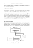
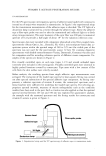
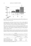
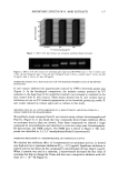
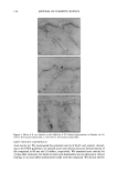
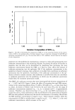
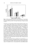
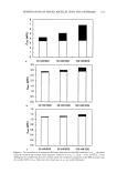
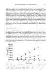
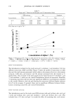
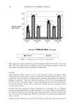
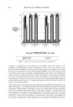
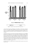
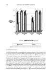
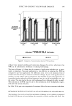
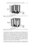
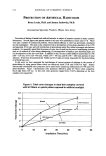
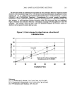
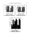
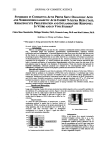
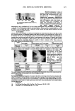
INHIBITORY EFFECTS OF R. A/JORI EXTRACTS 135 INHIBITION OF MELANOGENESIS B-16 melanoma cells were placed in a 25-ml T-flask at a density of 1 x 10⁵ cells/flask and cultured at 37øC in Dulbecco's Modified Eagle's Medium (DMEM, Sigma) con- taining 4.5 g/1 oFglucose, 10% (v/v) Fetal bovine serum (FBS), and 1% (v/v) antibiotic- antimycotic (Gibco, Auckland, N.Z.). AFter 24 hours of cultivation, we replaced the medium with new DMEM medium containing R. •orJ extracts of various concentra- tions. AFter five days, we washed the cells with phosphate-buffered saline (PBS) and collected the cells by trypsinization and centrifugation. We separated melanin From the pellet of the cells using 5% (w/v) trichloroacetic acid, dissolved the melanin in 1N NaOH solution, and checked the melanin content by absorbency at 475 nm. EVALUATION OF R. MORI EXTRACTS ON TYROSINE SYNTESIS IN B-16 MELANOMA CELLS (TYROSINASE ZYMOGRAPHY) Analysis of tyrosinase synthesis was performed by the modified method of Imokawa and Mishima (5). Detergent-solubilized cell extracts were subjected to SDS gel electropho- resis as Follows: Cell extracts (2 mg/ml protein) were electrophoresed on 10% (w/v) polyacrylamide gels. Total protein was measured by protein assay kit (Bio-Rad Labora- tories, California). AFter electrophoresis, the gel was placed in renaturation buffer [50 mM Tris-HCl (pH 8.0) and 2.5% (v/v) triton X-100] at room temperature For one hour. The gel was then incubated in developing buffer [0.1 M sodium phosphate (pH 6.8), 0.2% (w/v) L-DOPA] at 37øC For Four hours. Upon visualization of the tyrosinase bands, the gel was removed and dried, and the relative amount of tyrosinase band in each lane was quantified. EVALUATION OF R. MORI EXTRACTS ON TYROSINASE GENE EXPRESSION IN B-16 MELANOMA CELLS (RT-PCR) Total RNA was prepared using RNA PLUS TM (Quantum, Quevec, MW) From B-16 melanoma cells. Five micrograms per milliliter of total RNA was reverse transcribed by incubating the sample For one hour at 42øC in 25 pl of reaction mixture containing 200 U of MMLV (Moloney murine leukemia virus reverse transcriptase, Promega, Madison, WI) 1 pl of 100 pmol sequence specific primer dNTP (dATP, dCTP, dGTP, dTTP, Promega) and 1X buffer (50 mM Tris-Hcl, 75 mM KCI, 3 mM MgCI• and 10 mM DDT). Ten milliliters of RT reaction mixture was added to 40 pl of PCR mixture containing 1X PCR buffer (50 mM KC1, 10 mM, Tris-HC1 (pH 9.0), 1.5 mM, MgCI•, and 0.1% Triton X-100) 1 pl of 100 pmol Forward and reverse primer 5 pl of 2.5 mM each dNTP 2 pl of 25 mM MgCI• and Five units of Tag DNA polymerase (Promega). Amplification was performed at 33 cycles at 94øC For 30 sec, 50øC For 30 sec, and 72øC For 50 sec with the Gene AMP PCR system 2400 (Perkin Elmer, Oak Brook, IL). Two microliters of loading dye were added to 10 pl of amplification products, and the mixture was analyzed by 2% Agarose (Sigma) gel electrophoresis. EVALUATION OF R. RIORI EXTRACTS ACTIVITY ON MELANOGENESIS IN ANIMAL TEST R. mori extracts were dissolved at a final concentration, 1%, 5% (v/v) in dissolving solution (butylene glycol: H20 = 50:50). This solution was topically applied to sepa-
Purchased for the exclusive use of nofirst nolast (unknown)
From: SCC Media Library & Resource Center (library.scconline.org)