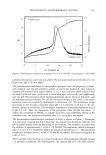
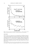
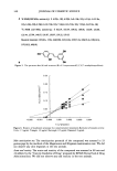
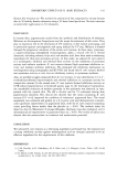
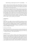
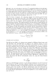
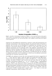
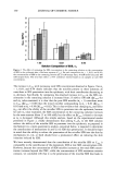
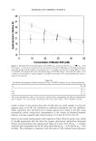
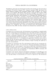
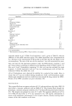
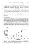
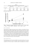
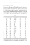
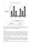
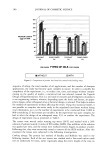
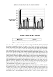
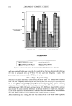
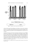
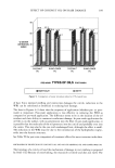
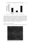
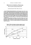
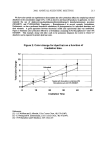
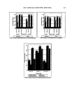
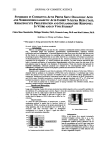
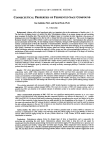
VITAMIN E ACETATE PENETRATION STUDIES 121 Table I Chemical Composition (INCI) of Three Cosmetic Formulations (LC, RP, and RS) Containing Vitamin E Acetate Product INCI LameIlar cream, LC (vitamin E acetate) Lameliar cream, RP (20% Roviparts ©) Lameliar cream, RS (20% Rovisome ©) Aqua, hexanediaol, cetearyl glucoside, Oenothera biennis, isopropyl is©stearate, behenyl alcohol, cetearyl isononanoate. glycerin, tocopheryl acetate, sotbit©l, dimethic©he, talc, sodium carhomer, tocopherol, hydrogenareal palm glycerides citrate Aqua, (Roviparts © E acetate), hexanediaol, cetearyl glucoside, Oenothera biennis, isopropyl is©stearate, behenyl alcohol, cetearyl isononanoate, glycerin, sotbit©l, dimethic©he, talc, sodium carhomer, tocopherol, hydrogenareal palm glycerides citrate Aqua, (Rovisome © E acetate), hexanediaol, cetearyl glucoside, Oenothera biennis, isopropyl is©stearate, behenyl alcohol, cetearyl isononanoate, glycerin, sotbit©l, dimethic©he, talc, sodium carhomer, tocopherol, hydrogenareal palm glycerides citrate Germany) and that contain vitamin E acetate (2.0%, in wt % active substance, Roche, Basel, Switzerland), along with controls, were tested for their pharmacokinetic proper- ties within the horny layer. The galenic formulations selected for this study exhibit physic©chemical similarities. The basic o/w cream (lameliar type, LC) contains 2% vitamin E acetate without any form of liposomes/microparticles. For the other two creams (RP, RS) the vitamin E acetate (1_0%) was packed either in Roviparts © (20%) or in Rovisome © (20%). The analytically determined content of vitamin E acrerate in the different creams was slightly lower, as shown in Table II. IRRITATION ASSAY IN THE IN VITRO SKIN MODEL (BUS) The isolated perfused bovine udder skin (BUS) model was introduced as an in vitro model to study skin penetration and irritation. Due to continuous perfusion, the horny layer demonstrates barrier and reservoir functions similar to those in the in vivo human situation. A comparison of the BUS and human skin stratum corneum by infrared spectroscopy was recently carried out (8). The natural skin model is widely used in dermat©logical and cosmetic research as well, and provides information concerning the penetration kinetics of active ingredients, their interaction in the skin, and skin com- patibility. Depending on the barrier function and the strength of the activant/irritant, increased arachidonic acid metabolism and a diminished cell viability can be analyzed biochemi- Table II Amount of Vitamin E Acetate (in wt%) in Different Lameliar Creams Determined by HPLC Products Analyzed in the product Analyzed on the strip Lamellar cream, LC (vitamin E acetate) Lameliar cream, RP (20 % Roviparts ©) Lameliar cream, RS (20 % Rovisome ©) 1.8 1.8 1.4 1.4 1.8 1.8

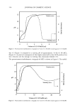
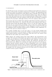
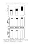
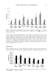
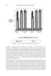
122 JOURNAL OF COSMETIC SCIENCE cally in whole skin biopsies. In determining the relevant part of the latter compound metabolism, the concentration of eicosanoids (ng PGE2/lag DNA) was measured in the skin (PGE 2 prostaglandin E2). The cell viability was assayed by the MTT assay (lag Formazan/lag DNA). (MTT methyl-thiazol-tetrazolium is a dye transformed by active mitochondria into a water non-soluble compound.) Details of the method concerning skin penetration and irritation are described elsewhere (9,10). APPLICATIONS For this investigation three independent udder studies were performed. The udders were perfused by an oxygenized and warmed-up Tyrode's solution. The skin surface tempera- ture was maintained at approximately 30øC. After maintaining the perfusion of the udder for a certain period in order to switch in an aerobic metabolism, the emulsions were applied topically with a surface density of 3-4 g/100 cm 2. The high dosage was intended to prevent any depletion of the vitamin concentration in the liposomal vesicles during the exposure period. Thirty minutes after starting the application, a dry paper towel carefully removed the residual cream. Whole skin punch biopsies with a diameter of D = 6 mm were taken 30 minutes after the end of the exposure to study the irritation potential of the creams applied. TAPE STRIPPING Adhesive tape stripping (Tesa R, 4204, BDF, Hamburg) was used to remove the outermost layers of the stratum corneum (SC) in sequence. Thirty and 90 minutes after the end of application, the adhesive tape strips for corneocyte layer removal were peeled off. Two parallel series of 15 successive strips (1.9 x 10 cm = 19 cm 2) were taken to analyze the vitamin E acetate content, either by the conventional HPLC method or UV spectroscopy. The chemical analysis for vitamin E acetate in either treated or untreated strips was performed in the Corporate Analytical Center of Henkel KGaA (D/_isseldorf, Germany). DETERMINATION OF VITAMIN E ACETATE BY HPLC For the determination of vitamin E acetate the adhesive tapes were extracted by means of acetonitrile. After solvent removal, the residue was dissolved in a well-defined volume of acetonitrile. This solution was injected in an HPLC system. The chromatographic investigation was performed using a reversed-phase HPLC column. Since vitamin E acetate shows fluorescence, this can be used for selective and highly sensitive detection. Application of an excitation wavelength of 276 nm and detection at 320 nm allows the quantification of vitamin E acetate as well as of underivatized vitamin E. The wave- lengths selected for excitation and detection do not induce fluorescence either of other ingredients of the cosmetic products or of skin constituents. The acquisition of a blank chromatogram is mandatory to ensure that any fluorescent components of the adhesive tapes are extracted. The limit of determination for standard injected samples is approxi- mately 5 ng.
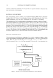
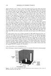
Purchased for the exclusive use of nofirst nolast (unknown)
From: SCC Media Library & Resource Center (library.scconline.org)