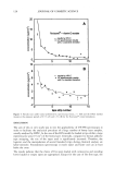
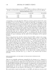
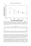
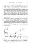
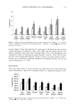
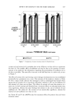
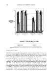
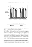
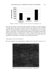
TOPICAL DELIVERY OF o•-TOCOPHEROL 165 TISSUE AND PREPARATION Micro-Yucatan pig skin obtained from Charles River Laboratories (Wilmington, MA) was used as the biological membrane to study in vitro percutaneous absorption. Upon receipt the freshly excised skin was washed gently with 1% (w/w) aqueous detergent, rinsed with deionized water, and patted dry with a paper towel. A 250-300-1am-thick layer of the skin was cut from the surface with a Padgett Electrodermatome TM instru- ment (Padgett Instrument, Kansas City, MO). The skin pieces were then rinsed and dried with paper towels before storage in plastic bags at 4øC. Skin was removed from the refrigerator and kept in isotonic solution to hydrate at room temperature one hour before starting the experiment. The dermatomed skin was cut into 10-mm circular pieces with a brass punch and placed epidermis-side-up in Bronaugh diffusion cells. The skin treated in this fashion from the stage of receipt until use retained its original permeability characteristics for four weeks after dermatoming (22). RADIOLABELING OF o•-T D-alpha-[3H]tocopherol was custom synthesized by Amersham Pharmacia Biotech, En- gland. This was received as a toluene:ethanol (9:1) solution with a specific activity of 19 Ci/mmol (molecular weight 432, at this specific activity). o•-T formulations were spiked with the radiolabeled o•-T such that each finite dose of 5-1al formulation applied on the skin contained approximately 300,000 dpm (disintegrations per minute). Gels were spiked before adding the gelling agent. Biphasic o/w formulations were spiked before addition of the aqueous phase. Analyses for the radiolabeled o•-T throughout this ex- periment were carried out with a liquid scintillation counter (LSC, Beckman Instru- ments). STATISTICAL DESIGN OF THE EXPERIMENT The application of formulations on the pig skin was carried out using a statistically approved model. A randomized complete block design was chosen as the design for the experiment. The statistical model appeared as shown in Table III. For the other studies, the application of each formulation was completely randomized over the number of cells and days of the study. DOSING Finite dosing was used to simulate the actual use conditions in all the in vitro permeation and metabolism experiments. The smallest volume of the formulation required, to obtain complete and uniform coverage of the diffusion cell surface area (0.636 cm2), was determined to be 5 lal, corresponding to a weight of about 4 mg for each formulation. After application, the preparation was uniformly spread on the stratum corneum (SC) side of the skin with the help of a glass rod, and the tip of the rod was washed into a vial containing 1.5 ml of ethanol in order to account for the material lost on spreading. With this technique, the exact amount of material applied to the skin surface was determined. To estimate the amount of o•-T in the original formulation, 5 lal of the

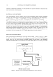
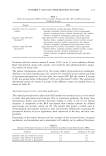
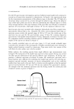
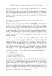
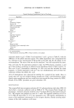
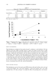
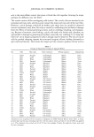
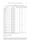
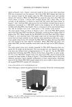
166 JOURNAL OF COSMETIC SCIENCE Table III Statistical Randomized Complete Block Design for the Application of Formulations Formulation Day s 1 Day 2 IPM solution W1 b Y1 d X1 c Z1 e Gel 1 W2 Y2 X2 Z2 Gel 2 W3 Y3 X3 Z3 Gel 3 W4 Y4 X4 Z4 Emulsion 1 W5 Y5 X5 Z5 Emulsion 2 W6 Y6 X6 Z6 Emulsion 3 W7 Y7 X7 Z7 Two consecutive days, 1 and 2. b,c Replicates on day 1 for IPM solution. d,e Replicates on day 2 for IPM solution. formulation was counted for its radioactive counts after equilibration of each formulation for a period of 24 hours. IN VITRO SKIN PERMEATION/METABOLISM METHODOLOGY A flow-through system was used for conducting in vitro permeation experiments. The total system consisted of a receptor fluid reservoir a variable flow rate peristaltic pump, Cassette © (Manostat, New York) a circulating water bath, Lauda © (Brickman Instru- ment, Westbury, NY) and a two-cell-holding heating block, 14 Teflon © flow-through diffusion cells, and a Retriever IV fraction collector (ISCO Inc., Lincoln, NE) to collect effluent fractions over the adjusted time period. Each diffusion cell had an inner diameter of 9 mm and a surface area of 0.636 cm 2 exposed to the receptor fluid. The receptor fluid was pumped at a flow rate of 1.5 ml/h from the reservoir to the diffusion cells placed in the holding blocks. The skin surface temperature was maintained at 32øC by adjusting the circulating water bath temperature to 39øC (23). The effluent from the diffusion cells was collected directly into glass scintillation vials every four hours for a period of 24 hours. SKIN TREATMENT The liquid scintillation counting technique was used to analyze all the in vitro perme- ation samples. In each experiment a minimum of four replicates were used. At the conclusion of the experiment, scintillation fluid was added to the effluent samples collected directly into the vials and the amount of ot-T penetrated was estimated from the counts of radioactivity present in the samples. Counts were obtained as dpm, which were then converted into micrograms of active by taking into account the spiking ratio for each formulation and specific activity of the active.
Purchased for the exclusive use of nofirst nolast (unknown)
From: SCC Media Library & Resource Center (library.scconline.org)