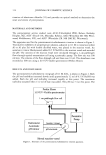
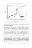
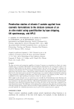
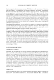
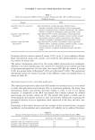
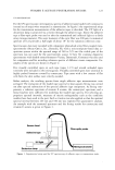
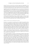
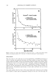
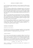
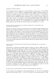
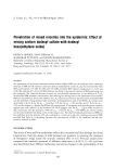
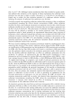
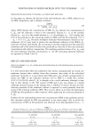
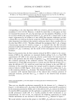
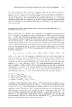
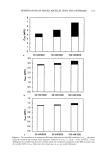
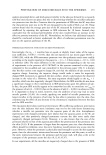
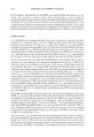
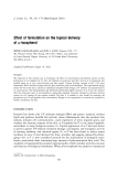
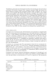
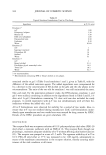
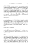
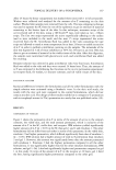
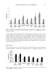
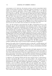
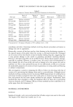
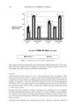
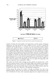
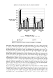
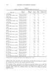
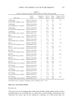
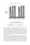
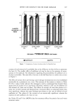
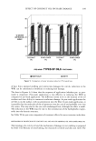
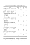
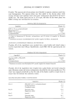
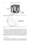
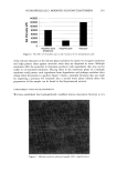
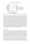
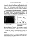
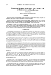
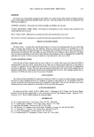
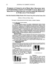
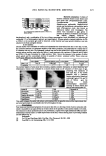
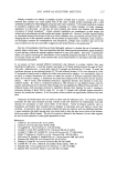
136 JOURNAL OF COSMETIC SCIENCE rated areas on the back skin of guinea pigs (n = 9) for two days (200 pl application, twice/day) before UVB radiation, while the dissolving solution alone was applied to the other area as a control. These areas were then irradiated once with 1,350 mJ/cm 2 using a UVB lamp (Vilber Loutmat, Marine La Vallee, France). After two weeks, the UV- irradiated site was stained by the Fontana-Masson staining method. SEPARATION AND IDENTIFICATION OF AN ACTIVE COMPOUND Separation. We extracted dried young twigs with 70% ethanol solution, using a vacuum rotary evaporator to concentrate the extract to dryness. To isolate the tyrosinase inhibitor from ethanol extract, we purified the extract through solvent fractionation, silica column chromatography, and Prep-LC. The ethanol extracts were dissolved in ethyl acetate, and then the residue was crystallized from CHC13. We purified the solid using silica chro- matography (Merck 200-400) and finally isolated the tyrosinase inhibitor. Identification. We crystallized the isolated tyrosinase inhibitor from ether benzene to yield pale yellowish prisms. The compound was identified by infrared (IR) spectroscopy, mass chromatography, and nuclear magnetic resonance (NMR). RESULTS INHIBITION OF TYROSINASE ACTIVITY AND MELANIN SYNTHESIS R. mori extracts were selected as potent tyrosinase inhibitors through our screening methods. We checked the extracts' inhibition activity on tyrosinase and melanin syn- thesis by changing the concentration (10, 20, 50, and 100 pg/ml). The extracts showed high tyrosinase inhibition activity. Melanin synthesis was also inhibited by R. mori extracts at a concentration of 50 pg/ml. The extracts showed no cytotoxicity. See Table I. EFFECT OF R. MORI EXTRACTS ON TYROSINE SYNTHESIS AND GENE EXPRESSION To examine the inhibitory mechanism of R. mori extracts on melanogenesis, we did the zymography for tyrosinase content in B-16 melanoma cells and RT-PCR for tyrosinase gene expression. Tyrosinase zymography showed that R. mori extracts at a concentration of 50-100 pg/ml did not lessen the tyrosinase synthesis (the band intensity does not change) (Figure 1). We also checked whether R. mori extracts inhibit the tyrosinase gene level by using RT-PCR. Figure 2 shows that the tyrosinase gene level was not changed by the treatment of R. mori extracts. These two results mean that R. mori extracts inhibit only tyrosinase activity, not tyrosinase synthesis and tyrosinase gene expression. Table I Inhibitory Effects of R. mori Extracts on Tyrosinase and Melanin Synthesis R. mori extracts (pg/ml) Tyrosinase inhibition (%) Inhibition of melanin synthesis (%) 10 56.0 13.5 20 65.8 18.5 50 89.5 36.5 100 97.8 65.3

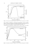
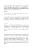
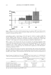
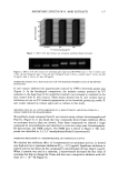
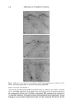
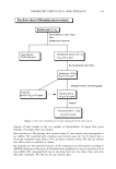
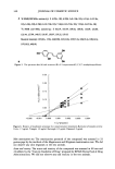
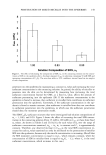
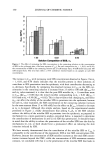
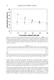
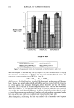
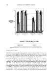
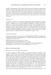
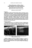
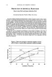
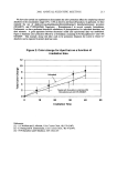
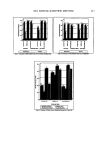
INHIBITORY EFFECTS OF R. MORI EXTRACTS 137 100,. • 80 • 60 • 40 • , 2o i 0 Control 50 1 O0 R. mori extracts(ug/ml) Figure 1. Effect of R. mori extracts on tyrosinase synthesis (band intensity) Figure 2. Effects of R. mori extracts on tyrosinase gene expression (RT-PCR). Lane 1' Tyr-•control. Lane 2: Tyr--R. mori 50 pg/rnl. Lane 3: Tyr--R. mori 100 pg/rnl. Lane 4: Actin--control. Lane 5: Actin--R. mori 50 pg/rnl. Lane 6: Actin--R. mori 100 pg/rnl. INHIBITORY EFFECT OF R. MORI EXTRACTS ON UVB-INT)UCED PIGMENTATION OF BROWNISH GUINEA PIGS R. mori extracts inhibited the pigmentation induced by UVB in brownish guinea pigs (Figure 3). In the histological comparisons, the melanin content produced by UV radiation in the basal layer of the epidermis (control) was increased as compared to the skin treated with R. mori extracts. These results showed that R. mori extracts had an inhibitory activity on UV-induced pigmentation in the brownish guinea pig model. R. mori extract induced no irritant signs such as redness in this study. IDENTIFICATION OF AN ACTIVE COMPOUND IN R. MORI EXTRACTS AND EVALUATION OF A SINGLE COMPOUND ON MELANOGENESIS We purified a single compound from R. mori extracts using column chromatography and Prep LC. (Figtire 4). It was found that four compounds showed high inhibitory effects on tyrosinase activity (data not shown). From these compounds we isolated a single compound by recrystallization and identified its structure using mass chromatography, IR spectroscopy, and NMR analysis. The NMR data is shown in Figtire 5. The com- pound was identified as 2,3',4,5'-tetrahydroxystilbene(2-oxyresveratrol). INHIBITION MECHANISM OF 2-OXYRESVERATROL ON TYROSINASE ACTIVITY We checked the inhibitory effect of 2-oxyresveratrol on tyrosinase activity. It showed very high activity in tyrosinase inhibition (IC5o = 0.23 pg/ml). Significant inhibition in enzyme activity was shown by this compound in concentrations of more than 0.1 pg/ml. When L-tyrosine was used as a substrate, 2-oxyresveratrol decreased the Km value of tyrosinase but did not change the Vmax, and thus was a competitive inhibitor with a Ki value of 5 x 10 -6 M (Figure 6).
Purchased for the exclusive use of nofirst nolast (unknown)
From: SCC Media Library & Resource Center (library.scconline.org)