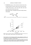
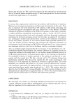
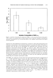
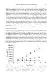
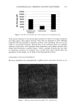
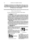
144 JOURNAL OF COSMETIC SCIENCE skin (1,2,9,17-20). Although various mechanisms have been invoked to explain surfac- tant-induced skin irritation, in the majority of these mechanisms the surfactant must penetrate into the skin in order to induce irritation (1,3,7,9,10,19-23). Accordingly, a simple way to reduce the skin irritation potential of a surfactant solution involves reducing the amount of surfactant that penetrates into the skin. A widely accepted view regarding surfactant-induced skin irritation is that, at surfactant concentrations exceeding the critical micelie concentration (CMC), where surfactant micelies first form, only surfactant monomers can penetrate into the skin, either because surfactant micelles are not surface-active or because they are too large to penetrate into the SC (3,6,14,16,18,24,25). This description of surfactant monomer penetration into the skin will be referred to hereafter as the monomer penetration model. The monomer penetration model is based primarily on experimental observations using mixtures of surfactants, where surfactant-induced skin irritation was correlated with the CMC of the surfactant mixtures examined (6,24,26). The surfactant monomer concentration is ap- proximately equal to the CMC (27), and therefore, according to the premise of the monomer penetration model, only the surfactant monomers should contribute to the observed surfactant-induced skin irritation. We have recently challenged the monomer penetration model by unambiguously dem- onstrating that micelles of the anionic surfactant sodium dodecyl sulfate (SDS) contrib- ute significantly to SDS penetration into the epidermis at SDS concentrations exceeding the CMC (28). The fact that SDS micelies were found to contribute to SDS penetration into the epidermis clearly contradicts the monomer penetration model, which predicts that the micellar surfactant should have no effect on surfactant penetration into the epidermis. In addition, we demonstrated that the SDS micelle contribution to skin penetration can be eliminated by mixing SDS with poly(ethylene oxide) (PEO), a non- ionic polymer known to bind to SDS micelies, to form PEO-bound SDS micelies (28). To explain both findings, we proposed a new model of surfactant penetration into the skin, in which the free SDS micelies are sufficiently small to access the aqueous pores of the SC, while the PEO-bound SDS micelles are sterically hindered from doing so due to their larger size. In contrast to the monomer penetration model, the new surfactant skin penetration model highlights the potential importance of the micelles in determining surfactant penetration into the skin. If the miceIlar surfactant is able to penetrate into the skin, then one predicts the commonly reported dose-dependent skin irritation re- sponse to surfactants (2,3,8,13,16,18), as well as providing an explanation for the increased penetration of surfactants into the skin beyond the CMC (25,28,29). The monomer penetration model fails to predict this observed dose dependence because at surfactant concentrations exceeding the CMC, where the concentration of surfactant monomers is constant, there should be no effect of increasing the total surfactant concentration on the surfactant-induced skin irritation. An important question that arose from our previous investigation (28) is whether mixing surfactants will have an effect on the ability of the micellar surfactant to penetrate into the skin. It is well known that mixing surfactants can lower the surfactant monomer concentration (24,30,31). In fact, the relationship observed between the reduction in the surfactant monomer concentration due to mixing surfactants and the resulting skin irritation reduction was used as the basis for the monomer penetration model (6,24,26). However, having demonstrated that the micellar surfactant can contribute to surfactant penetration into the skin (28), it became important to determine whether mixing

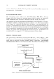
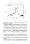
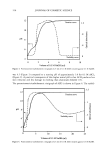
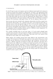
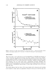
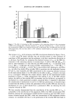
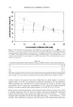
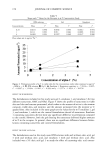
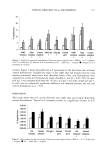
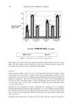
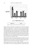
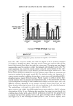
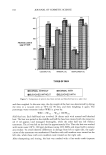
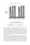
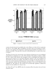
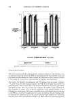
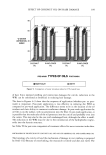
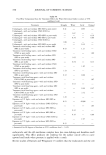
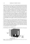
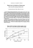
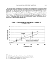
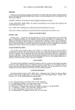
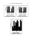
PENETRATION OF MIXED MICELLES INTO THE EPIDERMIS 145 surfactants could also reduce the penetration of the miceIlar surfactant into the skin in addition to reducing the surfactant monomer penetration. In this respect, it also became important to determine the relative contributions of the monomeric and the miceliar surfactant fractions to surfactant penetration into the skin, including quantifying which one is reduced the most by mixing surfactants. With this in mind, we measured the amount of SDS that penetrates into the epidermis from aqueous mixtures of SDS and the nonionic surfactant dodecyl hexa(ethylene oxide) (C12E6) after five hours of exposure. We found that SDS in SDS/C12E 6 mixed micelies is less able to penetrate into the epidermis than SDS in pure SDS micelies. We also found that the majority of SDS penetrating into the skin from SDS/C•2E 6 mixtures results from the monomeric fraction. Dynamic light scattering (DLS) measurements indicated that mixing SDS with C•2E 6 leads to an increase in the micelie size. We propose that it is the increased micelie size that reduces, or prevents, the penetration of the SDS/ C•2E 6 mixed micelies into the epidermis. Furthermore, we propose that, in general, surfactant mixtures that obey the monomer penetration model contain mixed micelies that are too large to be able to penetrate into the epidermis. EXPERIMENTAL MATERIALS Sodium dodecyl sulfate (SDS) and sodium chloride (NaCI) were purchased from Sigma Chemicals (St. Louis, MO) and were used as received. Dodecyl hexa(ethylene oxide) (C12E6) was purchased from Nikko Chemicals (Tokyo, Japan) and was used as received. Water was produced using a Millipore Academic water filter. •4C-radiolabeled SDS was purchased from American Radiolabeled Chemicals (St. Louis, MO) and was used as received. Phosphate-buffered saline (PBS) was prepared using PBS tablets from Sigma Chemicals and Millipore filtered water. PREPARATION OF SKIN SAMPLES Female Yorkshire pigs (40-45 kg) were purchased from local farms. Skin from the back of the pig was harvested within one hour of sacrificing the animal. The subcutaneous fat was trimmed off using a razor blade, and the full-thickness pig skin was cut into 2-cm x 2-cm pieces and stored in a -80 øC freezer until used. EXPERIMENTAL PROTOCOL After allowing the skin to thaw for a half hour at room temperature, the pig skin was mounted in a vertical Franz diffusion cell (Permegear Inc., Riegelsville, PA), with the SC facing the donor compartment. The donor and the receiver compartments of the diffusion cell were filled with phosphate-buffered saline (PBS phosphate concentration of 10 mM NaCI concentration of 137 mM Sigma Chemical Company), and the skin was left to hydrate for one hour. The PBS in the donor compartment was removed, and 1.5 ml of surfactant solution was added to the donor compartment. The solution in the donor compartment, referred to hereafter as the contacting solution, contained mixtures of
Purchased for the exclusive use of nofirst nolast (unknown)
From: SCC Media Library & Resource Center (library.scconline.org)