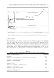
J. Cosmet. Sci., 64, 355–370 (September/October 2013) 355 Determination of physicochemical properties of delipidized hair ROGER L. McMULLEN, DONNA LAURA, SUSAN CHEN, DONALD KOELMEL, GUOJIN ZHANG, and TIMOTHY GILLECE, Materials Science Department, Corporate R&D, Ashland Specialty Ingredients, Wayne, NJ 07470. Accepted for publication April 22, 2013. Synopsis Using various physicochemical methods of analysis, we examined human hair in its virgin and delipidized state. Free lipids were removed by a solvent extraction technique (covalently bound lipids were not removed) using a series of solvents with varying polarity. We analyzed the surface properties of hair by conducting me- chanical combing and dynamic contact angle analysis. In addition, we used inverse gas chromatography surface energy analysis to explore the chemical composition of the hair surface based on interactions of various nonpolar and polar probes with biological molecules residing on the hair surface. Further, we investigated the importance that free lipids play in the internal structural properties of hair using dynamic scanning calorim- etry and tensile strength measurements. The microstructure of the hair surface was probed by atomic force mi- croscopy, whereas the lipid content of hair’s morphological components was determined by infrared spectroscopic imaging. We also monitored the water management properties of virgin and delipidized hair by dynamic vapor sorption, which yielded unique water sorption isotherms for each hair type. Using all these techniques, dif- ferences were found in the chemical composition and physical behavior of virgin and delipidized hair. To better understand the infl uence of hair lipid composition on hair styling treatments, we conducted mechani- cal analyses of hair shaped into omega loops to determine the stiffness, elasticity, and fl exibility of hair–polymer assemblies. Although there were no discernible differences between untreated virgin and delipidized hair, in terms of stiffness and elasticity, we found that treatment with hair styling agents produced different effects depending on the hair type used. Likewise, streaming potential measurements were carried out to monitor the binding capacity of rinse-off treatments on virgin and delipidized hair. Using this technique, we moni- tored the surface potential of hair and found signifi cant differences in the binding behavior of cationic poly- mers and surfactants (polyquaternium-55 and quaternium-26) on both hair types. INTRODUCTION Lipids play an important role in the functional and structural properties of hair. They are often designated as surface or internal lipids, and can be of sebaceous origin, or they may carry out a structural role. The structural lipids can be covalently bound [e.g., 18-methyleicosanoic acid (18-MEA) on the surface] or free (e.g., free fatty acids, cholesterol, ceramides, etc.). Address all correspondence to Roger McMullen at rmcmullen@ashland.com.

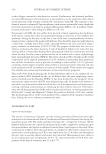
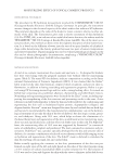
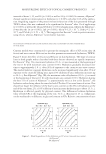
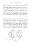
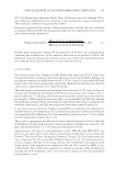
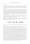
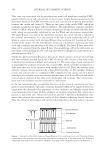
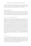
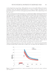
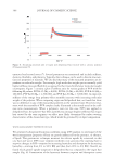
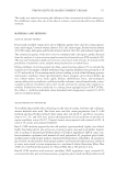
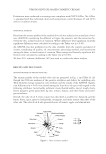
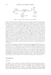
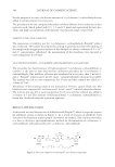
JOURNAL OF COSMETIC SCIENCE 356 They carry out a structural role by providing hair with a cell membrane complex (CMC), present in both cuticle and cortical cells. In recent years, insight has been gained as to the structural details of the CMC for both cuticle and cortical cells as well as the interface between the cuticle and cortex (1). There are two parts of the cuticle CMC, which are normally classifi ed as the upper and lower β layers. The upper β layer contains covalently- bound 18-MEA, which is believed to form a monolayer interspersed with other free fatty acids, which are presumably stabilized by van der Waals and electrostatic interactions. The upper β layer is located on the uppermost lamina of the cuticle cell and is exposed to the external environment. It is also present at the top of each underlying cuticle cell where it comes in contact with the lower β layer of an overlying cell. Located on the un- derside of the cuticle cell, the lower β layer consists of a monolayer containing free fatty acids and covalently attached fatty acids (but, no 18-MEA). The lower β layer of an over- lying cell is separated from the upper β layer of an underlying cell by the delta layer, in- tercellular cement holding the two cells together, which is thought to be glycoprotein or globular protein (Fig. 1). Unlike the upper and lower β layers in the cuticle, which contain a mixture of covalently and noncovalently attached lipids, the CMC of cortical cells consists of free fatty acids, cholesterol (or cholesterol sulfate), and ceramide (1). The outer edge of each cortical cell is surrounded by a bilayer structure, the cortical CMC, which is further enveloped by a delta layer that acts as the interface with another cortical cell. Thus, two bilayers from adjacent cortical cells are separated by a thin delta layer (Fig. 2). The interface between cuticle and cortical cells is a composite CMC composed on the cuticle side of a mixed monolayer of covalently and noncovalently attached fatty acids (lower β layer) and on the cortical side of a bilayer of free fatty acids, cholesterol, and ceramide. The cuticle mono- layer and cortical bilayer are also separated by a delta layer. In the last several decades, considerable attention in the research community has been given to structural lipids, especially covalently bound 18-MEA (2–4). Lipids of sebaceous origin were often deemed as less important, or even a nuisance, and thought to play little or no functional role in hair. In skin, sebum is an integral component that carries out a protective functional role. Sebaceous secretions provide a lipid-rich hydrophobic sub- stance that protects the outermost surface of skin, thereby enhancing barrier function. They also transport antioxidants and antimicrobial peptides to the skin surface to protect Figure 1. Schematic of the CMC (lower and upper β layers) of two overlying cuticle cells. The lower β layer corresponds to the overlying cell whereas the upper β layer corresponds to the underlying cell. Reprinted with permission from Society of Cosmetic Chemists. Copyright 2009. Originally appeared in Reference 1.
Purchased for the exclusive use of nofirst nolast (unknown)
From: SCC Media Library & Resource Center (library.scconline.org)