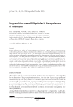
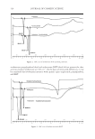
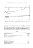
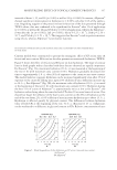
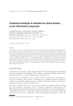
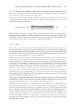
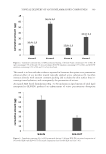
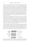
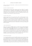
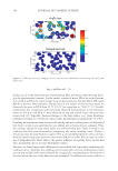
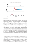
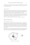
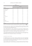
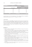
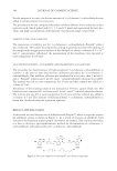
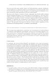
PHYSICOCHEMICAL PROPERTIES OF DELIPIDIZED HAIR 357 it from environmental insult (e.g., UV radiation) and invasion by foreign pathogens. Se- baceous lipids consist of free fatty acids, triglycerides, free cholesterol, cholesterol and wax esters, paraffi ns, and squalene. In this work, we investigated the physicochemical properties of delipidized hair using cutting-edge instrumental techniques to probe the surface and interior of the fi ber struc- ture. During our extraction procedure, we removed both sebaceous lipids and free struc- tural lipids from the exterior and interior of the fi ber. For this reason, it is diffi cult to discern differences in the structural/functional contributions of measured fi ber properties by either sebum or free structural lipids. It should be noted though that we do not re- move covalently attached lipids (e.g., 18-MEA) from hair. Therefore, any changes we observe in the physicochemical properties of delipidized hair by solvent extraction are due to free structural lipids or lipids of sebaceous origin. MATERIALS AND METHODS Studies were conducted on virgin and delipidized hair. We removed noncovalently at- tached lipids from hair using a Soxhlet extraction technique. Various instrumental tech- niques were used to examine differences between virgin and delipidized hair and to discover the importance of these lipids in the physicochemical behavior of hair. Details of each experimental technique are provided in the following sections. DELIPIDIZATION OF HAIR Using a Soxhlet extraction apparatus, free internal and surface lipids were removed from hair. This method is based on an established procedure in which hair is extracted with a series of solvents of increasing polarity (5). The apparatus consisted of a round bottom fl ask to which a Soxhlet extraction tube was mounted. Inside the Soxhlet extraction tube, a bundle of hair was placed in a cellulose thimble. A condenser was mounted on top of the Soxhlet extraction tube. The effect of solvent extraction on hair was investigated fi rst by treat- ment with t-butanol and n-hexane for 4 h each, then with a mixture of chloroform/methanol Figure 2. Schematic of the cortical CMC of two adjacent cortical cells separated by a delta layer. Reprinted with permission from Society of Cosmetic Chemists. Copyright 2009. Originally appeared in Reference 1.

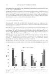
JOURNAL OF COSMETIC SCIENCE 358 (70:30, v/v) for 6 h. In each procedure, 3 g of hair was treated with 250 ml of solvent in the Soxhlet extractor. MECHANICAL COMBING MEASUREMENTS Combing analysis was achieved using a miniature tensile tester (Model 170), manufac- tured by Dia-Stron, Ltd., Hampshire, United Kingdom. The combing measurements were carried out on wet and dry hair with the following instrumental parameters: range, 2000 G gauge, 2 G size, 50 mm phase 1 (extension), 350% phase 2, 0% phase 3, 0% and phase 4, 0%. In all experiments, hair tresses were combed several times to remove entanglements before performing combing measurements. DYNAMIC CONTACT ANALYSIS A Thermo Cahn Dynamic Contact Angle Analyzer, DCA-322 (Thermo Fisher Scientifi c, Waltham, MA), was used to determine contact angle. A single dry hair fi ber, cut to ap- proximately 1 cm, was immersed in deionized water approximately 2–4 mm and the advancing contact angle was measured (inserted tip end fi rst). Fibers were obtained from the middle of a hair tress assembly ensuring that a straight section of fi ber was sampled. The required hair diameters were measured using a laser micrometer (Mitutoyo, model LSM-5000) purchased from Dia-Stron, Ltd. Twenty fi bers each for virgin and delipidized hair were measured. ATOMIC FORCE MICROSCOPY Atomic force microscopy (AFM) studies were conducted using a Digital Instruments Nanoprobe III Multimode SPM manufactured by Veeco Instruments (Santa Barbara, CA). Two modes of analysis, AFM and lateral force microscopy (LFM), were carried out in the contact mode. An AFM/LFM probe head was used in conjunction with a 100 μm piezoelectric scanner. The instrument used is a “beam bounce” type in which a diode laser beam is focused on to the back (top) of the cantilever. Commercial AFM probes (silicon tip on nitride lever, model SNL-10) were purchased from Veeco. Three-dimensional to- pography, error signal, and lateral force images were generated and analyzed with the instrument software (NanoScope Software, Veeco, Santa Barbara, CA). FTIR SPECTROSCOPIC IMAGING A 1-cm-long hair bundle was cut from the middle of Asian hair tresses and mounted on the top of a sample holder by embedding in ice. The hair bundle was then microtomed at -30°C into 5 μm thick cross sections with a Leica CM 1850 cryostat (Leica Microsystems, Inc., Bannockburn, IL). Hair cross sections were collected onto CaF2 windows for IR imaging. This preparation technique avoids any possibility of contamination with embedding or fi x- ing medium. Hair cross sections were imaged with a Perkin Elmer Spotlight system that couples an FTIR spectrometer with an optical microscope. The system consists of a linear
Purchased for the exclusive use of nofirst nolast (unknown)
From: SCC Media Library & Resource Center (library.scconline.org)