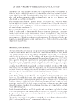
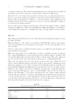
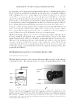
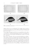
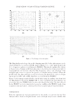
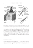
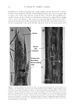
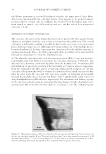
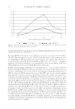
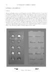
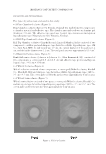
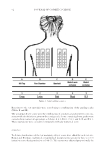
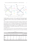
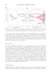
JOURNAL OF COSMETIC SCIENCE 22 using a pipette, then randomly cut with rapidly rotating poly(ethylene) blades in a manner similar to that mentioned previously (1). The resulting fragments were collected by means of a centrifuge, stained with a Giemsa’s solution (Wako Pure Chemical Industries, Osaka, Japan) (18), washed briefl y with a 50% (w/w) aqueous solution of glycerol, and then mounted to a microscope slide glass. A weight (30 g/cm2) was placed on the cover glass (grade: No. 1 Matsunami Glass, Osaka, Japan) while sealing the edges with Canada balsam. Mechanical slicing of a hair fi ber. Several strands of hair (cf16) were dried and embedded in an epoxy resin (Poly/Bed 812) according to the protocol of PolySciences (Warrington, PA). The solidifi ed resin block was sectioned to about 8 μm thickness by means of the microtome (American Optical, Buffalo, NY model 820) and stained with a 0.5% (w/v) aqueous solution of gentian violet. The microscope specimen was prepared in a man- ner similar to that described previously. MATERIAL FLOW PROPERTY OF MEDULLA H+ ion-fl ow through the medulla: the use of Congo red as an indicator. The hair fi bers (jf8, about 25 mm in length) were kept in an aqueous solution (5 mL) of 7 M urea, 2% (w/v) SDS, and 15% (v/v) ME at about 55°C for 20 min.1 The resulting fi bers were washed briefl y with water and dipped in a 0.025% (w/v) aqueous solution of Congo red (5 mL) for 1 d at ambient temperature. Three strands of the wholly stained hair were then placed, each in a U-letter shape, on the glass plate (22 × 22 × 0.17 mm) which had been glued with the epoxy resin to the window hole made in the bottom of a plastic Petrie chamber Figure 2. The o bservation chambers for the material fl ow study of the M. The type (A) was used for the in- verted microscope, whereas the type (B) was mainly employed for the upright microscope. 1 Treatments of hair and wool fi bers with the medium containing ME for long time at higher temperature was previously reported (19, 20). In “H+ ion-fl ow through the M: The use of Congo red as an indicator” process, the hair fi bers were softened by soaking in the presence of ME for a short time of period to wholly stain the fi ber body with Cong red. During the staining period, the swollen shafts were nearly brought back to the dimensions of the starting unheated wet fi bers. Although keratins, the main proteins of hair, might be structurally disturbed to some degree in the dyeing process, H+ ions was found to fl ow preferentially through the medulla. The “OH--fl ow through the M: the use of phenolphthalein as an indicator” process, on the other hand, did not use ME in the fl ow study of OH- ions.

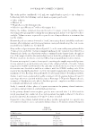
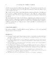
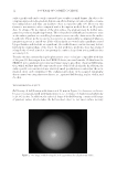
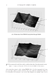
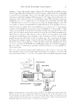
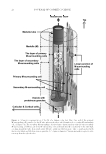
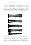
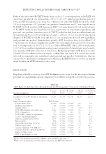
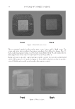
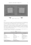
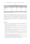
STRUCTURE OF HUMAN SCALP HAIR−II 23 (diameter 55 mm, bank height, 3 mm) cf. Figure 2B. Subsequently, the middle position of the U-shaped fi ber was cut with razor to give the two pieces which were hence antiparallel to each other on the glass plate. Next, each of the right- and left-end sections of the paired shafts was overlaid with a dampened blotting paper (5 × 15 mm), whereas the midsection (gap about 7 mm) of the fi bers was not covered with the paper. When the whole system was equilibrated to 25° ± 1°C, a glass lid (0.17 mm thickness) was temporally removed to add 0.5 M hydrochloric acid (0.05–0.1 mL) onto one of the two dampened paper covers. The color change occurring in the hair shafts was analyzed by means of the upright microscope. OH--fl ow through the medulla: The use of phenolphthalein as an indicator. A 0.5% (w/v) aqueous solution of phenolphthalein was put into a glass vial (diameter 22 mm, height 55 mm) with a screw cap till the solution level reached about 3 mm. Subsequently, the white hairs (jm67, about 40 mm in length) were vertically stood in the vial at ambient temperature, where the root side was in the indicator solution. After 7–14 d, the fi bers were briefl y washed with distilled water and extended on the aforementioned Petrie chamber in a parallel arrangement. Both sides of each of the fi bers were then covered with the damp- ened papers in a manner similar to that described in “H+ ion-fl ow through the medulla: the use of Congo red as an indicator.” When the whole system was equilibrated to about 25° ± 1°C, one drop of 1 M NaOH was carefully placed onto one of the blotting papers. The color change occurring in the hair shafts was followed by the use of the upright bio- logical microscope. Flow of the anthocyanin dye through medulla: Application of a DC voltage to a hair shaft. The white hair fi bers (jm67 about 30 mm length) were warmed in an aqueous solution of 8 M urea and 4% (w/v) SDS at 55°C for 2 h. One strand of the softened fi bers was put through Figure 3. The in strumental setup for the DC voltage-assisted fl ow of the purple anthocyanin dye. The white hair fi ber was put into a glass capillary, and the fi ber ends were dipped into the electrode solutions as de- picted in the fi gure. An aqueous solution of the cationic dye and the buffer solution were initially set as the positive and the minus electrode solutions, respectively see the procedure “Flow of the anthocyanin dye through M: Application of a DC voltage to a hair shaft” for more details.
Purchased for the exclusive use of nofirst nolast (unknown)
From: SCC Media Library & Resource Center (library.scconline.org)