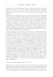
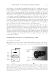
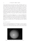
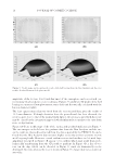
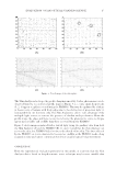
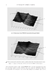
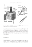
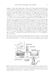
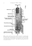
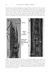
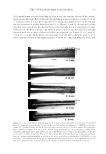
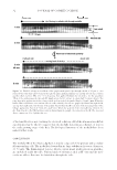
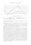
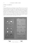
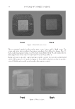
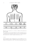
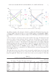
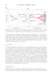
JOURNAL OF COSMETIC SCIENCE 24 the U-shaped glass capillary (inner width 1.0 mm, wall thickness 0.1 mm, and length 25 mm) which had been fi lled with pure water see Figure 3. After making each of the fi ber ends protrude by about 3 mm in length from the capillary terminals, the water in- side the capillary was soaked up with blotting paper, then the capillary ends were sealed with a poly(vinyl alcohol)-glue to prevent the infi ltration of the electrode solutions (vide infra) into the capillary. Subsequently, the capillary with the hair shaft was placed hori- zontally between positive and minus electric bathes [both: 2 cm (W), 2 cm (H), and 1 cm (D)], where the former and the latter bathes had been fi lled with a 0.5% (w/v) aqueous solution of the cationic anthocyanin dye and a pH 7/0.067 M PBS, respectively. Initially, the root side of the hair fi ber was put into the positive electric bath (anode). When the whole system equilibrated to about 25°C, DC 550 voltage, in most cases, was applied between the Pt-electrodes, using a power supply (model Crosspower 1000 ATTO, Tokyo, Japan) and an ammeter (model PC700 resolution, ±0.1 μA Sanwa, Tokyo, Japan). By means of the stereomicroscope, a dark purple-colored zone was recognized exclusively in the medulla and became longer toward the cathode side with increasing time of the voltage application. By reversing the electrode polarity, the colored zone was quickly moved back or faded out from the anode side, which had once been the cathode side. It was also noticed that a direct current was not only varied with the hair samples but gradually increased with time for instance, from 20 to 25 μA in one sample and from 1.3 to 2.5 μA in the other sample over a span of the experimental period (0–10 min). We consider that the current fl uctuation was caused by the uncontrollable electric conduc- tance over the wet hair surface and by the irregular inner structure of the medulla. Figure 4. The hai r fi ber (jf8) was treated with papain in pH 7/PBS at 30°C (the procedure “Enzymatic thin- ning of hair fi bers using the papain”). Pictures (A–F) were taken at the incubation time of 4 min, 2, 4, 6, 7, and 10 h, respectively. We consider on the basis of the diameter measurement that the dark part of the picture (E) is mainly composed of the M which consists of the M-surrounding cells and the tube cf. Figure 6.

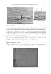
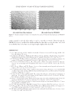
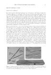
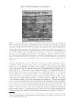
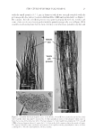
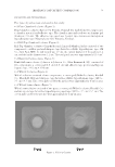
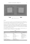
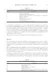
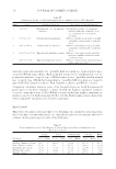
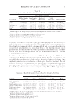
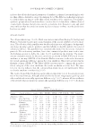
STRUCTURE OF HUMAN SCALP HAIR−II 25 RESULTS AND DISCUSSION MORPHOLOGY OF MEDULLA Recently, we reported the following microscopic observations on the cellular components of human scalp hairs (1) see Figure 1. The cuticular cell took a trowel-like shape, consist- ing of a handle-shaped part (CuH) and a blade-shaped part (CuB). The CuB parts over- lapped one another and partially fused together to form a layer (CuP) within the cuticular cell region. The cortical cell had a spindle-like shape and was similar in dimensions to CuH of the cuticular cell. The medulla, on the other hand, was appeared to possess a thin tubular wall. We, therefore, performed in the fi rst phase of the present study a detailed analysis of the medulla structure, using the aforementioned advantages of optical micros- copy. For this aim, the hair samples were either digested thin with papain or sliced with a microtome or randomly cut with a rotating cutter. Figure 4 shows the time course of the structural change of the young girl’s black hair fi ber (jf8) by an action of papain in the pH 7/PBS at 30°C. Although the inverted mi- croscope did not provide high-resolution images, the digestion of the hair fi ber seemed to take place in a phased manner. In other words, the outmost cuticular cell region was slowly stripped off from the hair shaft during the fi rst incubation period (about 5 h) Figure 4A–D. Then, the cortical cell region was digested within 2 h to provide the thin remainder which appeared to consist mainly of the medulla Figure 4E. The elders’ white hairs (jf65 and jm67) were digested in a similar fashion with the enzyme. The thin remainder such as the one seen in Figure 4E was useful for the structural study of the medulla. Namely, upon the mechanical cutting which was described in “Random mechanical cutting of enzymatically thinned hair fi bers,” two kinds of fl at and long substances (width 4–8 μm, length 70–90 μm, thickness 2–3 μm) were formed from the remainder as demonstrated in Figure 5. One of them is assigned as “a primary M-surrounding cell” and the other as “a secondary M-surrounding cell.” The primary M-surrounding Figure 5. The white ha ir fi ber (jf65) was treated with papain in pH 7/PBS at 30°C and then randomly cut Giemsa staining see the procedure “Random mechanical cutting of enzymatically thinned hair fi bers.” Panels (A and B) with bright fi eld illumination the panel (C) with oblique illumination.
Purchased for the exclusive use of nofirst nolast (unknown)
From: SCC Media Library & Resource Center (library.scconline.org)