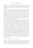
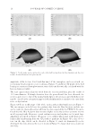
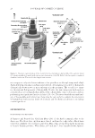
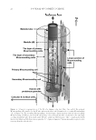
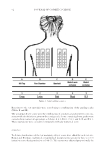
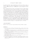
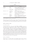
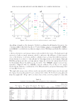
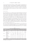
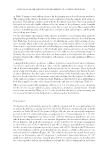
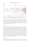
JOURNAL OF COSMETIC SCIENCE 26 Figure 6. Schematic represe ntation of the M of a human scalp hair fi ber. One end of the primary M-surrounding cell is attached to the M tube, whereas the other end is bound to the secondary M-surrounding cell cf. Figure 5A. The secondary M-surrounding cell also forms a linear junction with the other secondary M-surrounding cell. Hence, all of the M-surrounding cells are bound directly or indirectly to the tube wall, covering around the tube. In general, adults’ M tube, unlike an elderly persons’ tube, is neatly packed with the vesicles which are full of proteinous granules (0.5–1 μm in diameter). Various materials in aqueous solu- tion may fl ow through the tube.

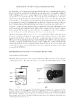
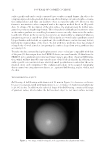
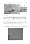
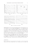
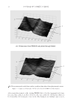
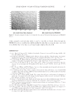
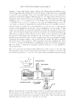
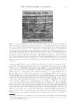
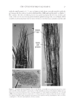
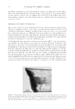
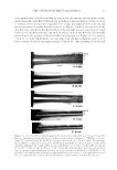
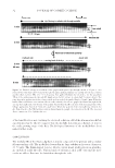
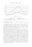
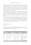
STRUCTURE OF HUMAN SCALP HAIR−II 27 cell was featured by the long thin fl at body see Figures 5A and 6. It appears that one end of the cell was attached on the surface of the medulla tube and the other end was linked to the secondary M-surrounding cell. In addition, a small irregular-shaped sub- stance, like plant roots, was usually observed on the primary M-surrounding cell see Figure 5A. It is considered that the irregular substance is a ripped off-product of the medulla tube (by the mechanical agitation), still binding partly to the primary M-surrounding cell. On the other hand, the secondary M-surrounding cell was character- ized by the long, fl at, and symmetric body with split ends Figure 5B and C. The cell was connected to the primary M-surrounding cell or to the other secondary M-surrounding cells as shown in Figure 5A see the linear junctions in Figure 6. The previous studies (9,11–16) on the hair structure did not mention the M-surrounding cells, presumably because the microscopic specimens were prepared by just cutting the hard hair fi bers by means of a microtome. We found that two kinds of the M-surrounding cells were very similar to each other in the terms of the dimensions and the staining susceptibility to gentian violet, Giemsa etc. therefore, these two kinds of cells are usually indistinguishable from each other, even under the polarized light illumination for instance, see Figure 7.2 Figure 7. A polarized light mi croscopic picture of the white hair fi ber (jm67). The medulla (M) appeared light brown for the most part. The regions of the M-surrounding cells, the cortical cells and the cuticular cells were seen in color yellow, light green and yellowish brown, respectively. The fi bers were warmed in a solution of 8 M urea and 4 wt.% SDS at 55°C for 2 h, washed successively with water and ethanol, sandwiched be- tween a slide glass and a cover glass using Canada balsam as a medium, then slightly pressed by means of a disk micrometer (CLM-DK Mitutoyo, Kanagawa, Japan) in a manner similar to that mentioned previously (1). Upon the compression, the softened fi ber (also swollen by about 1.3-fold of the original breadth) was fl attened at least in the width of about 100 μm hence, the retardation coloring due to an uneven thickness effect, if any, was minimized within the fl attened area. Although the angle between two nicols were properly adjusted to distinguish between the hair components in color, the primary M-surrounding cell region was not differentiated from the secondary one. The M tube appeared to be somewhat widened and fractured by the compression. 2 In o ur previous microscopic observations, the cortical cell region was not differentiable from the M-surrounding cells thereby, these regions were simply defi ned as a cortical cell region cf. the Figure 2 in the report (1) for the cross section of a human hair fi ber.
Purchased for the exclusive use of nofirst nolast (unknown)
From: SCC Media Library & Resource Center (library.scconline.org)