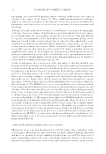
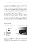
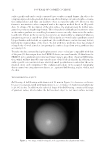
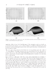
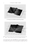
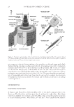
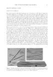
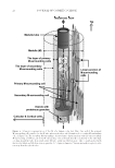
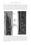
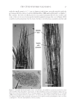
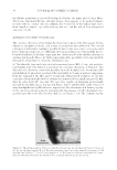
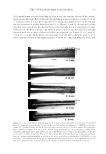
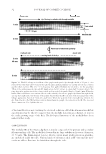
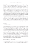
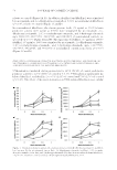
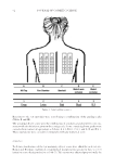
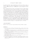
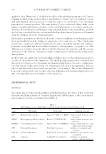
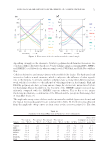
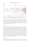
JOURNAL OF COSMETIC SCIENCE 28 In addition to the M-surrounding cells, a large tubular structure was found to exist in the M as shown in Figures 8A and 9A and B when the hairs were treated enzymatically to remove most of the outer substances from the fi bers. The M is thus regarded as the tubular substance which is doubly covered with the primary and secondary M-surrounding cells as depicted in Figure 6. We tentatively speculate that the M (tubular) wall is originated from the primary M-surrounding cells. Remarkably, many drum-shaped vesi- cles were arranged in a series inside the tube, and most of the vesicles were densely fi lled Figure 8. (A) The enzymatically t hinned hair fi ber consisting mainly of the M tube and the primary sur- rounding cells. The hair fi bers (cf16) were processed with papain in pH 7/PBS in a manner similar to that described in the procedure “Random mechanical cutting of enzymatically thinned hair fi bers.” The resulting substance, which corresponded to that seen in the panel E of Figure 4, was washed with water, cut by means of the poly (ethylene) blades, and stained with Giemsa solution. (B) The cross-sectional specimen of the hair fi ber (cf16 about 8 μm in thickness). The specimen was prepared by cutting along its longitudinal axis gentian violet staining see the procedure “Random mechanical cutting of enzymatically thinned hair fi bers.” The primary M-surrounding cell is not clearly distinguishable from the secondary one in this section of the fi ber shaft. M refers to the medulla.

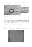
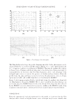
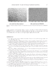
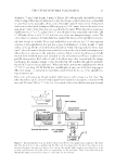
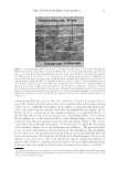
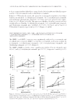
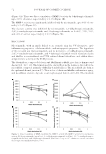
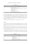
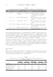
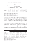
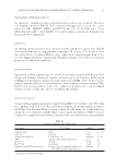
STRUCTURE OF HUMAN SCALP HAIR−II 29 with the small granules (0.5–1 μm in diameter) which was strongly stainable with the protein-specifi c dyes such as Coomassie Brilliant Blue (CBB) and gentian violet see Figure 9. By contrast, the tube of elderly persons was sparsely populated with the vesicles and some of the vesicles were loosely packed with the granules as typically seen in Figure 9B. It would be worth noting here that the hairs of a baby or an infant were generally very thin and Figure 9. (A and B) The drum-shaped ve sicles in the M tube (A and B) were pictured using the black hair (cf16) and the white hair (jm67), respectively. (C) The amorphous cluster of the granules which came ou from the vesicles of the white hair (jf65). The hair samples (3–5 mm length) were warmed at 30°C in a pH 7/PBS solution of papain (about 20 units) for 3–5 h, then in the fresh enzyme solution for another 30 min. The digested hair fi bers were randomly cut by means of the rapidly rotating poly(ethylene) cutter, and then centrifuged to collect the hair fragments Giemsa staining.
Purchased for the exclusive use of nofirst nolast (unknown)
From: SCC Media Library & Resource Center (library.scconline.org)