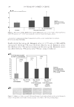
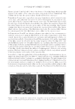
JOURNAL OF COSMETIC SCIENCE 178 The antihuman elafi n antibody (Abcam) was used at a concentration of 1/200 for one night at room temperature and amplifi ed with streptavidin/biotin. The technique involves three layers. The fi rst layer is an unlabeled primary antibody. The second layer is a biotinylated secondary antibody. The third layer is a complex of streptavidin–biotin peroxidase. The peroxidase (CliniSciences, Nanterre, France) produces purple-colored end products. They were observed using an optical microscope (Leica DMLB, Nanterre, France). Quantifi cation. The staining was detected with acquisition of a threshold that allowed selecting the staining in the interest area. The surfaces were then measured. The data were exported as values in an Excel fi le. Results and statistics. The quantifi cation of each band was relative to actin. The results were expressed in the protein percentage compared with the untreated control at 100%. Statistics were analyzed using a Mann–Whitney nonparametric test. IN VIVO CLINICAL TESTS Study design. The clinical study was carried out as a placebo-controlled double-blind randomized split-face study under dermatological control. The effi cacy of the formula containing Hamamelis at 1% was compared with the baseline (before treatment, D0) and to the other half-face treated with placebo. The study was conducted during a period of 56 d with check points at D0, D28, and D56 for the antiwrinkle effect, and during a period of 84 d with check points at D0 and D84 for the fi rming effect. This study was carried out in France from March to June 2013. Inclusion criteria. The study was performed on Caucasian female volunteers aged from 52 to 76 year, displaying wrinkles and fi ne lines on the crow’s feet of average to strong intensity and saggy skin on the face. Application modality. The products (containing Hamamelis at 1% or placebo formula) were applied by the volunteers twice a day, on each half face for 84 d. The application was carried out by the volunteers, by circular massages until complete penetration, especially on the crow’s feet area and the cheek. METHOD OF EVALUATION Measurement of skin roughness by fringe projection. Images were acquired on the crow’s feet area (surface of 12 cm2) via digital video camera coupled to a fringe projection system (Dermatop™ system, Breuckam, Meersburg, Germany, Eotech, Marcoussis, France) at D0, D28, and D56. ST and Stm parameters were calculated as follows (TopoSurf and Optocat analysis systems): - ST: maximum amplitude of the relief (mm). A decrease means a reduction of the main wrinkles. - Stm: mean difference b etween peaks and valleys (mm). A decrease means a smoothing of the studied surface. Measurement of the fi rmness of the skin with Dynaskin®. Dynaskin® (Orion Concept, Tours, France) is an add-on to the DermaTop™ system, which permits to evaluate the

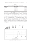
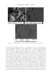
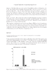
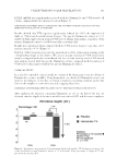
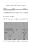
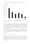
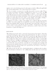
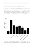
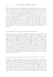
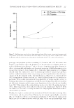
UNDERSTANDING SOLAR SKIN ELASTOSIS 179 fi rmness of the skin with a noncontact method. Dynaskin® produces a deformation close to the clinical approach by blowing air perpendicular to the area of interest or with a dedicated angle of 45°. The 3D sensor, using fringe projection techniques, captures just before the deformation of shape of the local surface and then when the deformation is applied. The parameters studied are the volume (mm3) and the maximal depth (mm) of the deformation. A decrease in these parameters shows an improvement in the fi rmness of the skin. Results and statistics. The results were expressed with the mean percentage of variation of the mean value of measured parameters: (meanDx-meanD0)/meanD0. The mean per- centages of variation compared with placebo were also calculated as mean variation of Hamamelis at 1% mean variation placebo. The normality of distribution was checked using Shapiro–Wilk test (level of signifi cance at 1%). The statistical comparisons of the evolution of the parameter with time and with the placebo have been performed with Student t-test (if the normality of distribution is confi rmed) or with the nonparametric Wilcoxon test or Mann–Whitney test (if the normality of distribution is rejected). The level of signifi cance is 5%. RESULTS TOO MUCH ELASTIN AND LACK OF LOXL1 UNDER UVA IRRADIATION IN THE DERMIS: AN INEFFICIENT BALANCE Using quantitative RT-PCR, we have shown in fi broblasts that under UVA radiation, the expression of elastin mRNA is increased by 7.5-fold, whereas the one of LOXL1 mRNA remains stable (Figure 1). This imbalance between the expression of elastin and LOXL1 suggests that elastin is not conveniently cross-linked and therefore that the amount of functional fi bers synthesized under UVA radiation is insuffi cient. Fi gure 1. Expression of LOXL1 and elastin mRNA in UVA-irradiated fi broblasts versus nonirradiated cells (0).
Purchased for the exclusive use of nofirst nolast (unknown)
From: SCC Media Library & Resource Center (library.scconline.org)