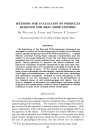
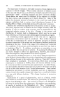
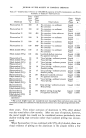
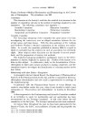
298 JOURNAL OF THE SOCIETY OF COSMETIC CHEMISTS The importance of melanin in the bodily economy of man depends on its capacity to absorb sunlight. When marked reduction of melanin occurs in the eyes and skin of man, as in albinism, the light-exposed skin becomes prematurely "aged": wrinkling, keratoses and even carcinoma develop. These effects are noted even in childhood if the exposure to sunlight has been intense and prolonged, as in South Africa (2). Also, in the albino the decreased amounts of melanin in the uveal tract and retinal pigment epithelium lead to serious visual disturbances because of the dazzling effects of light and marked decrease of visual acuity is common. Melanin also contributes significantly to the position of man in society. The newly acquired coat of melanin can be a badge of leisure, while the position of man in a given social structure may be determined by the congenital melanin content of his skin. Changes in the amount and distribution of melanin lead to disfigurement which may be no more than a blemish, or, if extensive and in deeply pigmented peoples, a tragedy. In considering normal pigmentation of the skin and its regulatory mechanisms, many factors must be considered. Mere surface inspection of the skin or examination of a section of epidermis may give an entirely erroneous impression of the actual rate and degree of melanin production obtaining at the time. Each of the stages to be outlined now is essential for completion of the process, and interruption at any level can lead to a breakdown (Fig. 1). In addition, acceleration or retardation at any level can affect the over-all rate of activity. In assessing the influence of any factor thought to affect skin pigmentation, therefore, due consider- ation must be given to the probable level at which it operates. The coloration of pigmented spots, or of the skin of Negroes and patients with Addison's disease reflects the activity of specialized cells, melanocytes these cells can be seen in the routine skin section as "clear cells" because their cytoplasm is vacuolated. It is known that, if fresh tissue containing melanocytes is incubated in DOPA, these cells become diffusely brown. It is also known that in certain blue-gray pigmentation (so-called Mongo- lian spot) bipolar dendritic melanocytes are located in the dermis and give rise to a blue color because of the Tyndall phenomenon. These dermal melanocytes are present in certain amphibians and here in response to various stimuli they rapidly change their shape from a small ball of black to a star with delicate dendrites. This change functions in protective coloration. Examination of a patch of pigmentation, such as a freckle, reveals that melanin is located outside the melanocyte, presumably in the cyto- plasm of the Malpighian cells. The melanin contained in the Malpighian cell must be derived from the melanocyte this requires some mechanism of transfer of melanin to the Malpighian cell. Thus, "melanogenesis" must be differentiated from "melanin pigmentation": me/anogenesis refers

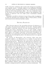
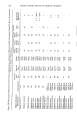
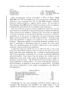
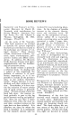
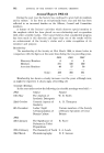
SOME ASPECTS OF MELANIN PIGMENTATION 299 . o ! Figure 1.--The epidermal melanin unit, from T. B. Fitzpatrick and A.D. Breathnach (3). N -- nucleus, M = mitrochondrion, PMS = pre-melanosome, MS -- melanosome, MG = melanin granule, G = Golgi membranes, E = endoplasmic reticulum and B.M. -- basement membrane. The development and transfer of pigmented melanin granules is illustrated in a series of five steps: I. Synthesis and condensation of the enzymic protein molecules. II. Arrangement of the molecules in structural form, leading to the development of an or- ganelle--the pre-melanosome. III. Biosynthesis of melanin and its accumulation within the pre-melanosome, proceeding through the stage of the melanosome to that of the fully synthesized melanin granule. IV. Transfer of melanin granules from melanocyte to Malpighian cells. V. Distribution of granules throughout successive epidermal levels and their final elim- ination at the surface as the result of progressive upward movement of cells from the basal layer to the stratum corneum. to the formation of melanin in the melanocyte, while melanin pigmen- tation describes not only formation of melanin but its distribution through- out the epidermal cells (Fig. 1). The dynamic process of melanin pigmentation involves two dissimilar cells (melanocyte and Malpighian cell) which appear to operate closely together as a single functioning unit each has an essential part to perform, and their roles are complementary. These two types of cells could be, considered to compose a structural and functional unit, an epidermal melanin unit (Fig. 1), analogous in some respects to other functional units such as the nephron. The epidermal melanin unit can be loosely defined as a melanocyte with an associated pool of Malpighian cells, the
Purchased for the exclusive use of nofirst nolast (unknown)
From: SCC Media Library & Resource Center (library.scconline.org)