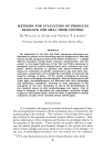
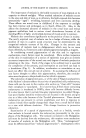
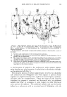
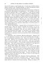
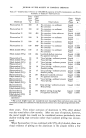
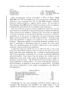
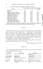
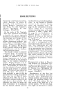
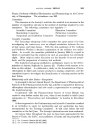
300 JOURNAL OF THE SOCIETY OF COSMETIC CHEMISTS size of which may be variable. Breathnach and Fitzpatrick (3) are not anxious to coin another term, but emphasis must be placed upon the role of the Malpighian cell as an active participant in melanin pigmentation and not upon its role as a purely passive recipient of melanin granules elaborated by the melanocyte. In fact, it is likely that the Malpighian cell may play an active role in controlling the synthesis of melanin granules by the melanocyte. After defining the epidermal melanin unit in so far as possible as a struc- tural unit, the respective roles of the individual components of this unit- (a) the melanocyte as the producer of melanin granules, and (b) the Mal- pighian cell as the vehicle of distribution throughout the epidermis----in the dynamic process of melanin pigmentation can now be examined. PRODUCTION OF MELANIN GRANULES IN MELANOCYTES In 194:9, when Lerner and Fitzpatrick (4:) were studying mammalian melanin formation, it became clear that the enzyme responsible for pro- duction of melanin was attached to particulates it was known that plant tyrosinases existed as soluble polypeptides. The specific nature of the particles was not investigated further at that time but about ten years later, Seiji, Bitbeck and Fitzpatrick (5) worked out the type and origin of • ,•o e Internal structure and TYROSINAS[ / TYROSIN [ • H ß { INTERMEDIATE COOH C•H Figure 2.--Melanogenesis in human skin, as seen in the light microscope, the electron microscope and at the molecular level. (From T. B. Fitzpatrick, M. Seiji and A.D. Mc- Gugan, New. Engl. J. Med., 265, 330, 1961). M = mitochondrion, PMS = pre-melanosome, MS = melanosome and MG = melanin granule.

SDME ASPECTS OF MELANIN PIGMENTATION 301 the particle that carries tyrosinase and is the subcellular site of melanin biosynthesis. Histochemical studies of human skin previously carried out at the Mayo Clinic by Fitzpatrick, Becker, Jr., Lerner and Montgomery (6) had shown that tyrosine formed dark brown particles about 0.2-0.5 • in diameter. It is now possible to give a better interpretation (Fig. 2) of the "DOPA" or tyrosinase reaction. 3lelanosorne (7) is the term given to the particle which is the subcellular unit of melanin biosynthesis. This particle is an organelle similar in some ways to a mitochondrion but distinct from a mitochondrion in fine struc- ture, chemical composition and development. The melanosome is not self-duplicating but undergoes a one-way progression: thus, new melano- somes do not arise from other melanosomes. The melanosome arises by a complex series of events, beginning in ribosomes, which are dense RNA- rich particles attached to the endoplasmic reticulum and the site of protein synthesis. TRANSFER OF MELANIN GRANULES FROM MELANOCYTE TO MALPIGHIAN CELL Until recently, it has been generally accepted that, upon leaving the melanocyte, mature melanin granules were (a) liberated into intracellular space and phagocytized as individual particles, or (b) in some way injected into the Malpighian cells. This transfer of particles from one cell to another was termed a "cytocrine" process by Masson (8). Studies with the electron microscope by Bitbeck in London (9) and Drochmans in Belgium (10) have shown that in hair and epidermis segments of dendrites containing melanin granules penetrate into Malpighian cells and are nipped off. It is impossible to say to what extent the penetration of dendrites that leads to this mechanism of transfer can be regarded as a dynamic activity on the part of the melanocyte or as a manifestation of phagocytosis by the Malpighian cell. Possibly the Malpighian cell plays a more active part in the transfer of melanin granules than was thought previously the Malpighian cell would be in a position to control the rate of transfer, stepping it up or slowing it down according to its requirements at a particular time. Such a process would be bound to influence the rate of melanosome synthesis. It seems probable that the rate of melanosome synthesis is geared to the rate of elimination and that the melanocyte as a secretory gland enters a resting stage unless it is called upon to produce melanosomes. For example, retinal pigment cells (in adults) are non- secretory glands and contain fully melanized melanosomes. Thus a sort of biological positive "feed-back" mechanism may exist in which, as the
Purchased for the exclusive use of nofirst nolast (unknown)
From: SCC Media Library & Resource Center (library.scconline.org)