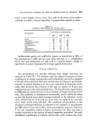
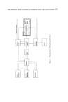
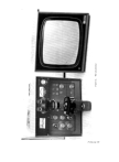
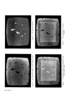
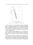
688 JOURNAL OF THE SOCIETY OF COSMETIC CHEMISTS which exhibit a wide range of sizes, only part of this range will be covered at any single magnification. This state of affairs arises when examining pigments, and introduces an element of the unknown into an evaluation of the complete size distribution. CONCLUSION It has been found that the Quantimet can be used to evaluate particle size with considerable advantage. The instrument is limited to the evalu- ation of particles which provide sufficient visual density, and by the morphology of the sample. Both free powders and suspensions of powders in transparent liquids and solids have been successfully examined. The use of this equipment has shown considerable practical advantages its use has removed a large part of the tedium of microscopical size analysis, as well as completely eliminating the subjectivity of size evaluation. Con- siderable time can, therefore, be saved, but the degree to which this is done is limited by the manual procedures involved (adjustment to present the sample to the microscope) and the number of particles to be counted (the time taken by the instrument is independent of total particle numbers whereas the time of the manual counting procedure is directly proportional to this number). It is unlikely that the capabilities of this instrument will be much im- proved in the near future. Alternative software is available for use with the analogue computer. Various means are available to present the image to the television camera including a microscope, an automated microscope, an electron microscope and an epidiascope (7, 17). Output from the system is available as meter read-out, or punched card or tape. Current developments of image analysis suggest that the next logical step in automatic image analysis must await the commercial appearance of an image system tied to a digital computer. Such a configuration is already under development (18), and promises a potential improvement in pattern recognition facilities. ACKNOWLEDGEMENTS The author wishes to thank Mr. A. Hooker of the South East Essex Technical College, Barking, who produced the micro-image of the test chart, and Dr. J. D. Lewis of the Rocket Propulsion Establishment, for permission to reproduce Fig. 11. (Received: 15th January 1969)

THE PARTICLE SIZE ANALYSIS OF PIGMENTS X¾ITH THE Q UANTIJ•IET 689 REFERENCES (1) Analyst 80 156 (1963). (2) Allen, T., Particle Size 2kleasurement (1968) (Chapman & Hall). (3) Irani, R. R. and Callis, C. F. Particle Size: Measurement Interpretation and Application (1963) (Wiley, New York). (4) Brit. J. Appl. Phys. õ (Suppl. no. 3) (1954). (5) British Standard 3406: Part 4 (1963). (6) A•talysis by microscopical methods for particle size distribution ASTM: E20-51T (1951). (7) Metals Research Ltd., I•Ielbourn, Roysion, Hefts. (8) Quantitative Television Microscopy, Micro-67, Microscope lO (April 1968). (9) Cole, M. Microscope and Crystal Front, •õ 148 (1966). (10) Fisher, C. Particle Size Analysis 77 (1967). (11) Kaye, B. H. Paint Oil Colour J. 1õ4 372 (1968). (12) Collins, G. F. Particle Size Analysis 65 (1967). (13) Smith, M. J. Microscope 10 123 (1968). (14) Herden, G. Small Particle Statistics (1960) (Butterworths, London). (15) Eckot•, R. K. The Microscope and Crystal Front 14 490 (1965). (16) Harwood, 5I. G. Brit. J. Appl. Phys. suppl. $, • S193 (1954). (17) Strang, A. Microscope 10 181 (1968). (18) Mendelsohn, M. L. et al. Advances in Optical and Electron 3Iicroscopy 9, (1968). (Academic Press, New York). (19) Franklin, A. G., Iron Steel Inst. 9,0 181 (1959). Introduction by the lecturer As one might expect, there are both advantages and disadvantages to be en- countered when using the Quantimet system. The former include rapid and relatively painless sample evaluation, while the principal disadvantage is usually presented by the nature of the specimen. Not all types of sample are suitable for evaluation by means of this technique although, provided a suitable optical image can be produced, useful results can be obtained. This is shown by the wide range of materials which are currently being examined with the instrument. In terms of the image presented to the instrument, the count is effectively absolute, i.e. if there is a particle seen on the screen, then the instrument counts it. The limitations to this result from the size, and the contrast and focus of the features while they are being examined. If the particle is partly out of focus in the microscope system, then it may or may not be counted, depending on how dense it is. In prac- tical terms, however, the results must be regarded' as relative, due to the variations which arise during sampling. Thus, emphasis should be placed upon establishing reproducable differences between two specimens, rather than upon a constant value for any single specimen. With the assistance of the instrument, sampling as a problem becomes much more obvious and much more easily solved. On the one hand we have more time to examine variations arising from different sampling techniques (unclouded by observer errors), while, on the other hand, we are able to examine greater numbers of particles in order to reduce the level of the error. It is interesting to note at this stage that we have found increased sampling levels to be useful in two distinct ways. Not only does it reduce the uncertainty of an evaluation (and hence the difference between two
Purchased for the exclusive use of nofirst nolast (unknown)
From: SCC Media Library & Resource Center (library.scconline.org)