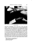
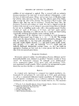
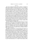
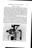
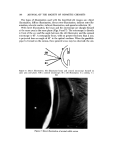
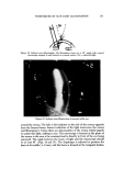
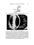
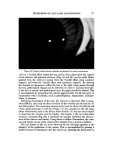
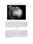
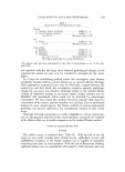
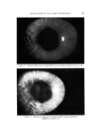
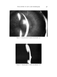
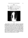
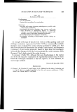
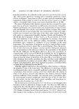
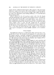
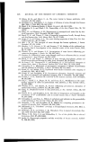
TECHNIQUES OF SLIT-LAMP ILLUMINATION 171 ß •.T. Figure 10. In'direct retro-illumination. Slit illuminator beam set at 45 ø angle with corneal microscope tangent to and focused on corneal surface. RL ----- reflected light Figure 11. Indirect retro-illumination of normal rabbit eye around the cornea. The halo is the brightest on the side of the cornea opposite h'om the focused beam. Internal reflection of the light transverses the cornea and illuminates it. Unless there are abnormalities of the cornea which impede or scatter this light, nothing is seen. The microscope is focused at the plane of the cornea in the area to be examined and is directly in h'ont of the eye being examined. The angle between the beam of light and the microscope should be at '_,east 45 ø (Figs. 14 and 15). The diaphragm is adjusted to produce the desirod slit width (1-2 mm) and the beam is focused at the temporal limbus.

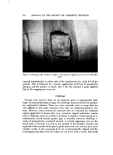
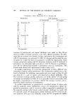
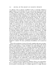
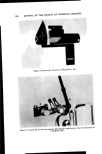
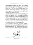
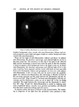
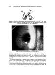
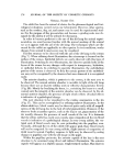
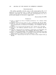
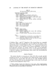
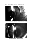
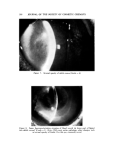
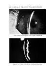
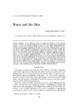
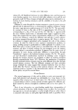
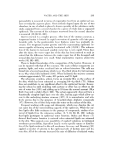
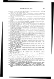
172 JOURNAL OF THE SOCIETY OF COSMETIC CHEMISTS $I •Cl't . Figure 12. Direct retro-illumination. Slit filuminator beam set at 45 ø with a 90 ø angle between corneal microscope, focused on cornea, and slit illuminator. Observer looks parallel to reflected light rays. RL • reflected light Figure 13. Direct retro-illumination of normal rabbit eye Sclerotic scatter illumination aids in detecting very slight and early changes in the corneal tissue which only mildly obstruct, but significantly scatter, light. Central circular clouding of the cornea is probably best determined by this method. In indirect illumination, a narrow beam is focused on nontransparent, trans- lucent tissue such as sclera, iris, or leukoma of the cornea. The microscope is focused at the same plane, slightly to the side of the beam. The angle between the corneal microscope and the slit illuminator is 60 ø or more, thus increasing the amount of internal reflection and the amount of light scattered (Figs. 16
Purchased for the exclusive use of nofirst nolast (unknown)
From: SCC Media Library & Resource Center (library.scconline.org)