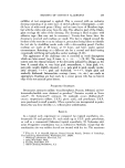
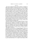
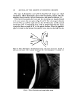
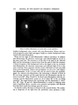
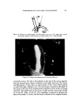
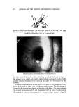
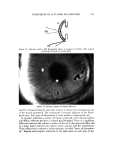
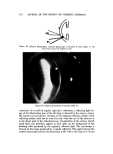
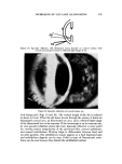
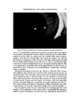
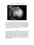
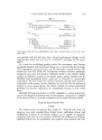
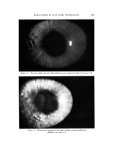
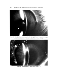
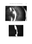
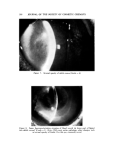
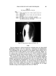
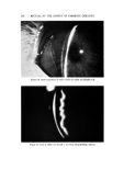
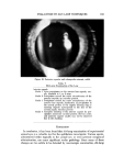
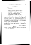
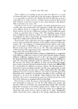
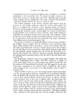
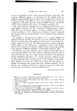
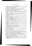
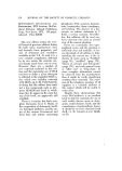
TECHNIQUES OF SLIT-LAMP ILLUMINATION 179 Figure 23. Fundus of normal rabbit eye spread evenly in the precorneal film and cover the entire surface of the cor- nea. A cotton-tipped swab dipped in 2% fluorescein and lightly applied to the upper conjunctiva, followed by eye-manipulated blinking, is required. Using diffuse illumination with a blue cobalt filter, the observer checks for epithelial defects (Fig. 9). An epithelial defect is present when fluorescein penetrates an area in which the squamous epithelial cells have been removed and stain the living cells of the stroma. It is not uncommon for an infrequent slight cot- heal staining to be observed in a rabbit population. SUMMARY The preceding discussion has centered around the utilization of the slit lamp in experimental situations. Ocular pharmacological and toxicological evaluations of pharmaceutical, cosmetic, and other formulations under inves- tigation in the experimental animals are easily and routinely made with the slit lamp. Experience in the past has demonstrated that the slit-lamp biomi- croscope has become an indispensable tool in aiding in investigations. The different types of illumination used in the clinical situation can be easily adapted to the experimental situation, and as one becomes familiar with the types of illumination, they can be utilized in routine examinations of the ex- perimental animal eye.

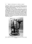
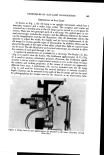
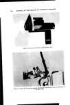
180 JOUBNAL OF THE SOCIETY OF COSMETIC CHEMISTS ACKNOWLEDGMENTS The authors gratefully note the review of this manuscript by Drs. A. R. Borgmann, O. J. Lorenzetti, and C. A. Robb. The permission of slit lamp man- ufacturers (Carl Zeiss, Inc., Bausch and Lomb, and Haig-Streit, Inc.) to re- produce photographs of instruments is appreciated. ( Received June 20, 1972 ) I•EFERENCES (1) Vogt, A., Lebrbruch und Atlas der Spaltlampenmikroskopie, Vol. 3, Springer, Berlin, 1931, pp. 1-227. (2) Goldmann, H., Biomicroscopy of the eye, Amer. J. Ophthalmol., 66, 789-812 (1968). (3) Hruby, K., Ueber eine wesentliche Vereinfachungder Untersuchimgstechnik des hin- tereu augenabschniltes im Lichthusche] der Spa]tlampe, Arch. Ophthalmol., 143, 224- 9 (1941). (4) Goldmann, H., An improved slit lamp apparatus, Brit. J. Ph!/siol., 13, 77-88 (1939). (5) Moses, R. A., The Goldmann app]anation tonometer, Amer. J. Ophthalmol., 46, 865- 9 (1958). (6) Jaeger, W., Tiefenmessung der menschlichen vorderlcanemer mir planparal]elen (Zu- satzgerat zur sp]atlampe), Graefe Arch. Ophthabno[., 153, 120-31 (1952). (7) Brown, N., Slit-image photography, Trans. Ophtha[mo[. Soc. UK, 89, 397-405 (1969). (8) Braley, A. E., Watzke, R. C., Allen, L., and Frazier, O., Sterescopic Atlas of Slit-Lamp Biomicro,copy, C. V. Mosby Co., St. Louis, 1970, pp. 1-115. (9) Berliner, M. L., Biomicroscopy of the Eye, Vol. 1, Cassel, London, England, 1949, pp. 100-26.
Purchased for the exclusive use of nofirst nolast (unknown)
From: SCC Media Library & Resource Center (library.scconline.org)