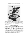
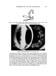
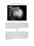
J. Soc. Cosmet. Chem., 24, 181-195 (March 2, 1973) Slit-Lamp Examination of Experimental Animal Eyes. II. Grading Scales and Photographic Evaluation of Induced Pathological Conditions It. A. BALDWIN, B.S.,* T. O. McDONALD, M.S.,* and C. H. BEASLEY, M.D.* Presented May 25, 1972, Seminar, Los Angeles, Calif. Synopsis-The SLIT LAMP has been shown to be a valuable tool for the examination of experimental animal EYES. With use of various techniques previously described, subtle as well as gross PATHOLOGICAL CHANGES in experimental eyes may be easily identified. These ocular pathological changes can be quantRated with numerical GRADING systems as outlined in this manuscript. These subjective grades center around the cornea, anterior chamber, iris, and lens. Ocular pathological changes can be documented by PHOTO- GRAPHING with the aid of a photoslit lamp. The use of stereophotography can be most useful because it gives the illusion of depth as one normally sees with the stereoptic vision of the slit lamp. INTRODUCTION Throughout the years various chemical entities have been studied in ani- mals prior to becoming either a useful therapeutic medicament or one of many thousands of compounds found to be too toxic or ineffective for use in humans. Animal models have also been devised in order to study similarities and differences between the natural or induced disease state and similar dis- * Science and Technology Division, Alcon Laboratories, Inc., P.O. Box 1959, Fort Worth, Tex. 76101. l' 1212 West Presidio, Fort Worth, Tex. 181

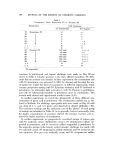
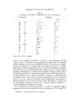
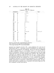
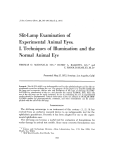
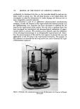
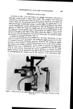
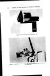
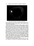
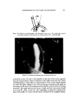
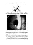
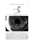
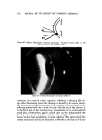
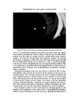
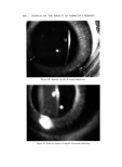
189, JOURNAL OF THE SOCIETY OF COSMETIC CHEMISTS eases in man. Through very carefully controlled experiments in animals, in- vestigators have gained a tremendous amount of knowledge which has been ultimately useful in the care and treatment of diseases in man. In the area of ophthahnology, animal test models are also an integral part of the study of disease processes of the eye. In this laboratory, in addition to the study of mechanisms of conditions similar to clinical eye diseases, each new therapeutic drug entity or cosmetic proposed for human use is carefully eval- uated in animals as an extra measure of consumer protection. And, as the scope of regulatory requirements broadens, other agents not specifically de- signed for direct ocular application but for periocular use must now be evalu- ated for effects on this sensitive organ. Ophthalmic, dermatologic, and cosmet- ic formulations are routinely evaluated in our laboratories for their potential to induce ocular damage in animal species prior to use in man. The use of the slit lamp as an aid in examination allows the investigator to make fine distinc- tions of ocular pathology. Thus, the investigator is able to make better judg- ments for selection of a formulation which possesses a low potential for ocular irritation. Of primary concern, regardless of the categories of interest, is that the in- vestigator is ultimately faced with documentation and quantitation of the experimental results. Of course, the animal investigator usually has an option not available to the clinicJan of terminal histopathologic ocular examination. Indeed, this is an extremely valuable tool but is not always optimal for follow- ing the course of a disease process. Therefore, appropriate methods for fol- lowing the disease and recording changes must be developed to evaluate in situ pathology. OCULAR IRRITATION GRADING SCALES In 1944, Draize and others (1) devised a system for evaluating ocular le- sions induced by topical applications of test chemicals (Table I). The Draize scale has thus become the mainstay for the evaluation of ocular lesions in ex- perimental animals and has been widely utilized as the basis for devising oth- er grading systems. Basically, the Draize scale is a subjective macroscopic evaluation of the eonjunetiva, cornea, and iris. Although originally intended for use in albino rabbits, modifications will allow use in other animal species and in specialized situations. As an outgrowth of the original Draize scoring procedure, our group as well as others have adopted the use of the slit lamp to the routine procedures of examination of experimental animal eyes. The slit lamp, developed by Gullstrand in 1911, was originally an exclusive re- search device which has now evolved to an indispensable tool for the ophthal- mie practitioner. We have found that this instrument is also an invaluable aid for the examination of experimental animal eyes in that gross as well as mi- nute pathological changes may be identified and subjectively quantitated in an in situ situation. With the development of the photographic capabilities
Purchased for the exclusive use of nofirst nolast (unknown)
From: SCC Media Library & Resource Center (library.scconline.org)