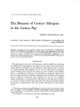
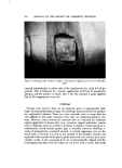
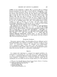
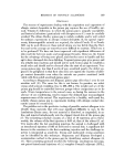
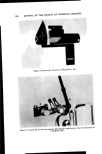
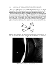
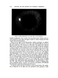
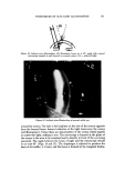
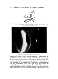
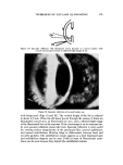
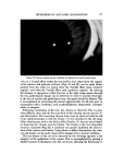
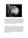
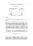
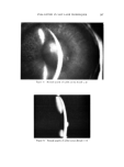
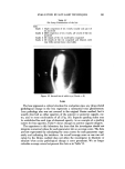
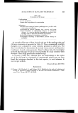
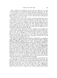
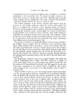
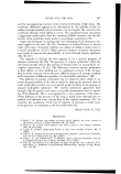
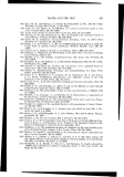
TECHNIQUES OF SLIT-LAMP ILLUMINATION 177 Figure 20. Normal rabbit anterior chamber as detected by small conical beam will see a Tyndall effect within the lens and by close observation the capsule of the anterior and posterior surfaces (Figs. 21 and 22) can be easily distin- guished from the cortex by noting when the Tyndall effect starts (anterior capsule) and when the Tyndall effect ends (posterior capsule). By noticing the changes of opaqueness within the lens as the light image passes through the lens, pathological changes can be detected. In order to examine thorough- ly the lens of normal and pathological eyes, the pupil should be dilated. This is accomplished by pretreating the animal approximately 15-30 min prior to examination with a mydriatic, such as phenylephrine, tropicamide, cyclopen- tolate, or atropine. Following examination of the lens, the vitreous is observed. This is some- what difficult, since only the first one-third of the vitreous can be seen by di- rect illumination. The remaining vitreous body must be observed with the aid of the ophthalmoscope or with the Hruby (3) lens attached to the slit lamp. Other attachments, such as the Braley-Allen Fundus (8) lens are most helpful in observing the vitreous and the fundus. The fundus is the last intraocular structure examined (Fig. 23). A mydriatic eye greatly facilitates the observa- tions of the vitreous and fundus. Using direct or diffuse illumination, the vitre- ous and fundus can be easily observed for changes from a normal condition. The last feature of the eye to be observed by the slit-lamp operator is the integrity of the epithelium of the cornea. This is accomplished by placing a limited amount of fluorescein into the cul-de-sac, allowing the fluorescein to

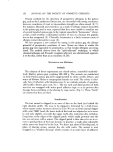
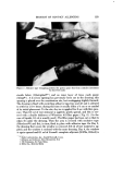
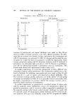
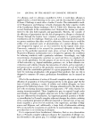
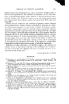
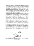
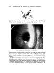
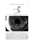
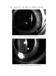
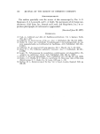
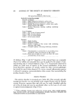
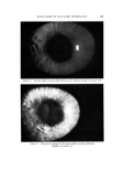
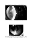
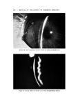
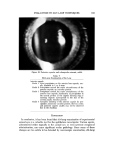
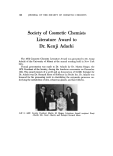
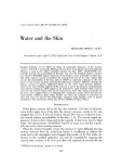
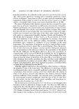
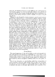
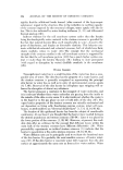
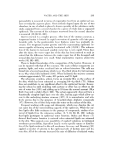
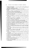
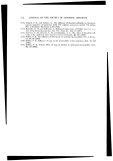
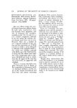
178 JOUBNAL OF THE SOCIETY OF COSMETIC CHEMISTS ... ß ... . ....,. .:..• ...•,::. ..... , .. • •.•ti•..•..-:.:: 1.5 i , ß ':f ? ß • ......-- :...• ./' ? • .:-.• .:• .• :.. .: •.. .. ...::.:•.•.. . . :.•:*-3_ ß ß , •." ...•:':•? •.•'•..:'4' :.:'.•.. ß ...'•.u3 . .. •.•.• Figure 21. Anterior capsule of normal rabbit lens Figure 22. Posterior surface of capsule of normal rabbit lens
Purchased for the exclusive use of nofirst nolast (unknown)
From: SCC Media Library & Resource Center (library.scconline.org)