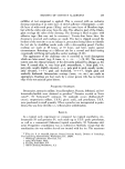
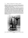
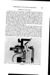
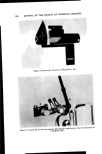
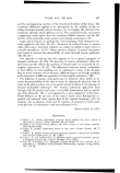
EVALUATION BY SLIT-LAMP TECHNIQUES 183 Table I Draize Scale" for Scoring Ocular Lesions _ Cornea A. Opacity--degree of density ............. Grades 0- 4 B. Area of cornea involved ................ Grades 1 -- 4 A X B X 5 Maximum score -- 80 Iris A. Values .............................. Grades 0- 9, A X 5 Maximum score -- 10 Conjunctiva A. Redness ............................. Grades 0- 3 B. Chemosis ............................ Grades 0- 4 C. Discharge ........................... Grades 0- 3 (A q- B q- C) X 9, Maximum score -- 9,0 The total score for the eye • sum ot• all scores øThe Draize scale has been abbreviated in this table. Consult Draize et al. (1) for com- plete table. now possible with the slit lamp, these induced pathological changes in the experimental animal eye may now be recorded in stereopsis by the inves- tigator. As a basis for establishing grading scales, the investigator must become completely familiar with the normal animal eye as viewed with the slit lamp. Then appropriate numerical scores may be arbitrarily assigned between the normal eye and that which the investigator considers maximal pathologic change for any given test situation. Although similar to the original Draize method of subjective scoring, as previously stated, minute changes may be identified and quantitated which could not be detected by macroscopic examination. We have found this method extremely valuable with regard to examination of the cornea, anterior &hamher, iris, and lens. Due to anatomical features in many animal species, the Draize method of scoring conjunctival pathology has proved satisfactory for quantitating reaction in this ocular tissue. Although slit-lamp examination is readily adaptable to various animal spe- cies, for the purposes intended in this communication, comments are confined to the albino rabbit eye to enable comparison to the original Draize method. STUDY OF ALBINO i•ABBIT EYE Cornea The rabbit cornea is examined first (Table II). With the aid of the slit lamp one may easily visualize three distinct layers: epithelium, stroma, and endothelium. Contrary to the Draize method, one is capable of separately examining each layer in most instances. With the aid of fluorescein staining, epithelial defects may be quantitated with regard to both intensity and area

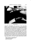
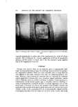
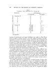
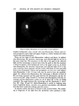
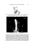
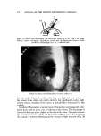
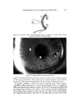
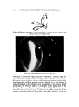
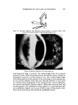
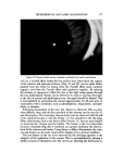
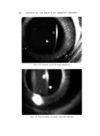
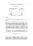
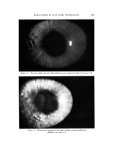
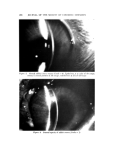
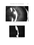
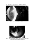
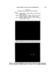
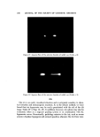
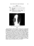
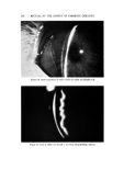
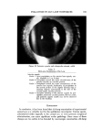
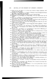
184 JOURNAL OF THE SOCIETY OF COSMETIC CHEMISTS Table II Slit-Lamp Examinations of the Cornea •pithelial staining (fluorescein) Intensity of staining Grade 1 Slight fluorescein staining in a given area Grade 2 Moderate fluorescein staining in a given area Grade 3 Marked fluorescein staining in a given area, under- lying structures still visible Grade 4 Extreme fluorescein staining, masking underlying structures Area of staining Grade 1 25% or less of cornea stained Grade 2 50% of cornea stained Grade 3 75% of cornea stained Grade 4 All of cornea stained Opacities Grade Grade Grade Grade 1 Slight clouding 2 Moderate clouding 3 Marked clouding of the cornea with underlying structures still visible 4 Complete opacity, obscuring underlying structures Panus Grade i Vascularization is present but is not invading from around the entire circumference of the cornea Grade 9• Panus has invaded 2 or more mm from the entire circumference of the cornea of staining (Figs. 1 and 2).* Opacities of the stromal layer are commonly observed in rabbits and generally these may be graded by the degree of stro- real clouding and/or corncal thickness (Figs. 3-7). Occasionally observed in rabbits are small areas of opacity in the corneal endothelium. It has been found that these opacities are usually masked by other corneal pathologies but should be noted when observed. If a severe cornea] inflammatory process is allowed to progress, neovascularization often occurs, thus necessitating the grading of panus (Fig. 8 ). Anterior Chamber The anterior chamber is examined next (Table III ). This normally optically transpm'ent chamber was not included in the original Draize scale for obvious reasons. However, aqueous flare and cells may be easily identified with the aid of the slit lamp and may be quantitated either on the basis of cell count or optical density judgments (Figs. 9-12). * Ocular pathologic changes were induced by various means. These include immtlllOo logic reactions (uveitis and corneal transplantation), formalin, detergents, shampoos, ancl various preservatives at concentrations to cause toxicity.
Purchased for the exclusive use of nofirst nolast (unknown)
From: SCC Media Library & Resource Center (library.scconline.org)