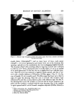
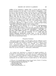
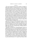
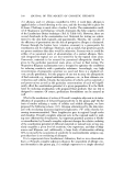
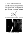
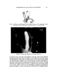
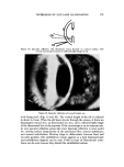
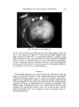
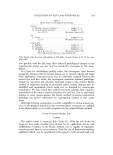
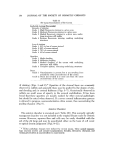
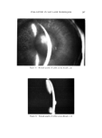
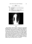
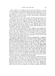
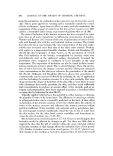
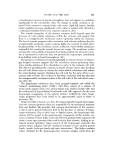
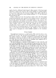
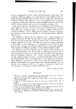
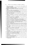
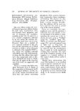
EVALUATION BY SLIT-LAMP TECHNIQUES 189 Table III Slit-Lamp Examination of the Anterior Chamber Flare Grade I Slight change in optical density from normal barely detectable flare Grade 2 Easily detectable turbidity within the anterior chamber moderate change in optical density Grade 3 Marked clouding of aqueous humor underlying structures plainly visible Figure 9. Normal anterior chamber of rabbit eye (Grade: 0) Figure 10. Aqueous flare (indicative of the presence of cells and/or protein material) in the anterior chamber of rabbit eye (Grade = 1). (Note: Flare is a Tyndall phenomenon)

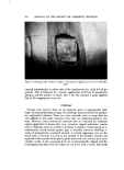
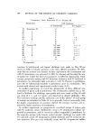
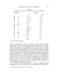
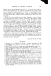
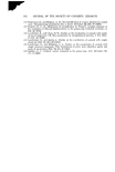
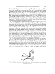
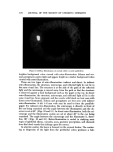
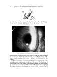
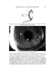
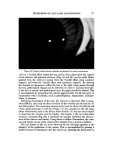
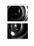
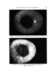
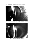
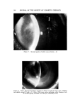
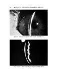
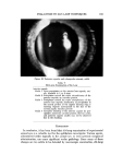
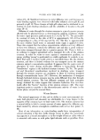
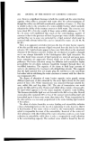
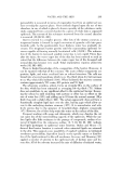
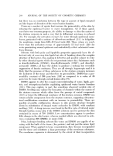
190 JOURNAL OF THE SOCIETY OF COSMETIC CHEMISTS Figure 11. Aqueous flare of the anterior chamber of rabbit eye (Grade = 2) Figure 12. Aqueous flare of the anterior chainbet of rabbit eye (Grade = 3) The iris is an easily visualized structure and is extremely sensitive to chem- ical irritation and immunogenic reactions. As in the Draize method, we have found that iris hyperemia may be easily quantitated with the aid of the slit lamp (Table IV) (Figs. 13-15). In addition, however, iris edema may also be evaluated, either as a separate grading system or in combination with the hyperemia scores. Occasionally, pathology anterior to the iris, such as severe anterior chamber hypopyon and corneal opacities, obscures the iris from view.
Purchased for the exclusive use of nofirst nolast (unknown)
From: SCC Media Library & Resource Center (library.scconline.org)