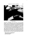
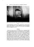
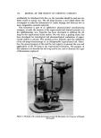
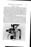
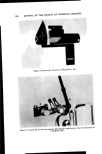
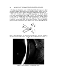
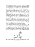
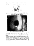
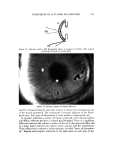
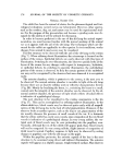
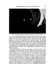
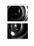
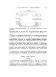
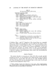
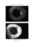
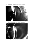
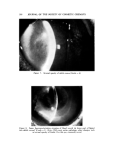
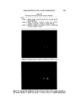
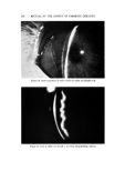
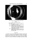
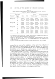
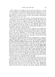
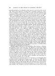
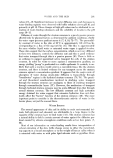
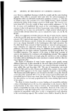
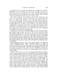
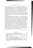
176 JOURNAL OF THE SOCIETY OF COSMETIC CHEMISTS NORMAL RABBIT EYE The rabbit has been the animal of choice for the pharmacological and toxi- cological evaluations carried out in our laboratories. However, other species, such as the monkey, dog, rat, and mouse may be used for experimental stud- ies. For the purpose of this presentation and because a grading scale was de- signed for the rabbit, it will be utilized for discussion. In order to become proficient in the use of the slit lamp for animal experi- mentation, one must become familiar with the normal anatomy of the animal eye as it appears with the aid of the slit lamp. The techniques which are dis- cussed for the rabbit are applicable to other species. In test conditions, ocular changes from normal to most severe may be visualized. The first structure to be observed with the aid of the slit lamp is the cornea (Fig. 7). When utilizing direct illumination, the microscope is focused on the surface of the cornea. Epithelial defects are easily observed with this type of illumination. Switching to retro-illumination, the observer quickly looks at the layers of the cornea for any changes with respect to transparency, hydration, or epithelial defects. In switching to specular illumination, the endothelium pattern of the cornea is observed. At first, the mosaic pattern of the endotheli- um may not be recognized by the observer but once observed it is recognized easily. The anterior chamber, which is posterior to the cornea, is the next area to be observed. The normal anterior chamber is invisible to light of the slit lamp because there are no discrete cellular units or reflective characteristics therein (Fig. 20). Merely by localizing the beam, i.e., restricting the beam to a small, conical unit, the integrity of the anterior chamber can be observed. In the ab- normal anterior chamber, the presence of light which reflects (Tyndall effect) is indicative of a pathological change. The next structure to be visualized in the routine examination is the iris (Fig. 17). This can be accomplished by utilizing indirect illumination. In the albino rabbit eye, blood vessels may be observed quite easily with all magnifi- cations of the slit lamp due to the lack of iridal pigmentation. However, if the observer looks at another species, such as the dog or cat, pigmentation of the iris usually obscures the presence of small vessels of the iris. It is fortunate that the albino rabbit has easily seen vessels, since congestion of the iris blood vessels is indicative of a pathological change. In very young rabbits, the ves- tigial ends of blood vessels may be seen protruding into the pupillary area. Normally, although the iris of the rabbit will appear pink, these blood vessels will not be congested. In very young rabbits, slight amount of congestion of iridal vessel is normal. Pupillary response to light may be observed by noting changes in pupillary size with the slit image on the pupil. Within the pupillary perimeter, the anterior capsule of the lens is the next structure to be examined using direct and indirect illumination. The anterior capsule will appear as a slightly opaque, rough-textured surface. The observer

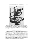
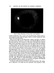
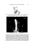
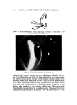
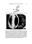
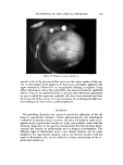
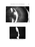
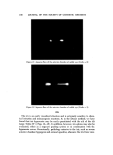
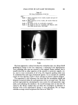
TECHNIQUES OF SLIT-LAMP ILLUMINATION 177 Figure 20. Normal rabbit anterior chamber as detected by small conical beam will see a Tyndall effect within the lens and by close observation the capsule of the anterior and posterior surfaces (Figs. 21 and 22) can be easily distin- guished from the cortex by noting when the Tyndall effect starts (anterior capsule) and when the Tyndall effect ends (posterior capsule). By noticing the changes of opaqueness within the lens as the light image passes through the lens, pathological changes can be detected. In order to examine thorough- ly the lens of normal and pathological eyes, the pupil should be dilated. This is accomplished by pretreating the animal approximately 15-30 min prior to examination with a mydriatic, such as phenylephrine, tropicamide, cyclopen- tolate, or atropine. Following examination of the lens, the vitreous is observed. This is some- what difficult, since only the first one-third of the vitreous can be seen by di- rect illumination. The remaining vitreous body must be observed with the aid of the ophthalmoscope or with the Hruby (3) lens attached to the slit lamp. Other attachments, such as the Braley-Allen Fundus (8) lens are most helpful in observing the vitreous and the fundus. The fundus is the last intraocular structure examined (Fig. 23). A mydriatic eye greatly facilitates the observa- tions of the vitreous and fundus. Using direct or diffuse illumination, the vitre- ous and fundus can be easily observed for changes from a normal condition. The last feature of the eye to be observed by the slit-lamp operator is the integrity of the epithelium of the cornea. This is accomplished by placing a limited amount of fluorescein into the cul-de-sac, allowing the fluorescein to
Purchased for the exclusive use of nofirst nolast (unknown)
From: SCC Media Library & Resource Center (library.scconline.org)