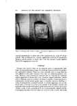
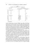
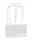
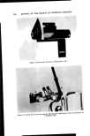
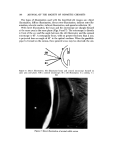
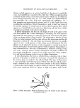
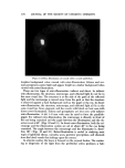
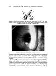
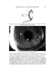
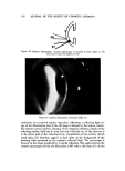
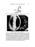
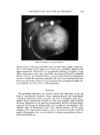
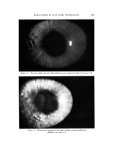
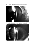
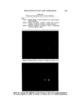
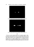
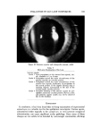
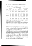
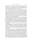
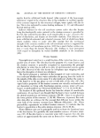
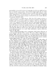
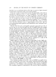
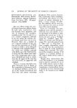
184 JOURNAL OF THE SOCIETY OF COSMETIC CHEMISTS Table II Slit-Lamp Examinations of the Cornea •pithelial staining (fluorescein) Intensity of staining Grade 1 Slight fluorescein staining in a given area Grade 2 Moderate fluorescein staining in a given area Grade 3 Marked fluorescein staining in a given area, under- lying structures still visible Grade 4 Extreme fluorescein staining, masking underlying structures Area of staining Grade 1 25% or less of cornea stained Grade 2 50% of cornea stained Grade 3 75% of cornea stained Grade 4 All of cornea stained Opacities Grade Grade Grade Grade 1 Slight clouding 2 Moderate clouding 3 Marked clouding of the cornea with underlying structures still visible 4 Complete opacity, obscuring underlying structures Panus Grade i Vascularization is present but is not invading from around the entire circumference of the cornea Grade 9• Panus has invaded 2 or more mm from the entire circumference of the cornea of staining (Figs. 1 and 2).* Opacities of the stromal layer are commonly observed in rabbits and generally these may be graded by the degree of stro- real clouding and/or corncal thickness (Figs. 3-7). Occasionally observed in rabbits are small areas of opacity in the corneal endothelium. It has been found that these opacities are usually masked by other corneal pathologies but should be noted when observed. If a severe cornea] inflammatory process is allowed to progress, neovascularization often occurs, thus necessitating the grading of panus (Fig. 8 ). Anterior Chamber The anterior chamber is examined next (Table III ). This normally optically transpm'ent chamber was not included in the original Draize scale for obvious reasons. However, aqueous flare and cells may be easily identified with the aid of the slit lamp and may be quantitated either on the basis of cell count or optical density judgments (Figs. 9-12). * Ocular pathologic changes were induced by various means. These include immtlllOo logic reactions (uveitis and corneal transplantation), formalin, detergents, shampoos, ancl various preservatives at concentrations to cause toxicity.

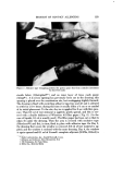
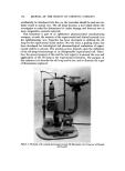
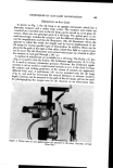
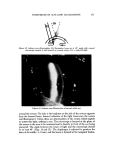
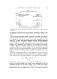
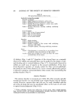
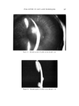
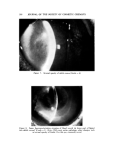
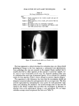
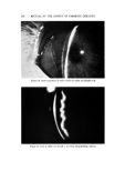
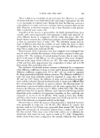
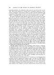
EVALUATION BY SLIT-LAMP TECHNIQUES 185 Figure I. Normal rabbit corneal epithelial fluorescein staining (Grade -- 0 area = 0) Figure 2. Fluorescein staining of damaged rabbit corneal epithelium (Grade -- 2 area: 1)
Purchased for the exclusive use of nofirst nolast (unknown)
From: SCC Media Library & Resource Center (library.scconline.org)