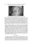
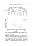
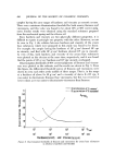
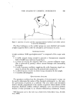
474 JOURNAL OF THE SOCIETY OF COSMETIC CHEMISTS In our laboratory, the flanks and back o[ the Mexican hairless dog have been observed to contain pigmented keratin [ollicular plugs which share some or certain clinical and histologic similarities to the comedones seen in man (9). In an endeavor to determine the suitability ooe these animals for routine acne experiments, a study was conducted to evaluate preparations currently employed to treat clinical acne. MATERIALS AND METHODS Four test sites, approximately 2.5 cm in diameter, with grossly evident follicular plugs, were selected on the back of each dog. Areas were carefully photographed prior to treatment to record control appearance and again at the conclusion of the study. Efficacy and irritation potential were evaluated from the resulting slides. Before and after treatinent, biopsies were obtained with a 6-mm Keyes cutaneous punch for histological evaluations. Following fixation of the tissues in 10% formalin, slides were prepared by routine histo- logic means, stained with hematoxylin and eosin, and examined utilizing stan- dard dermatopathologic criteria (10, 11) employed in recognizing inflamma- tory, degenerative, or proliferative changes. Test or control materials in solution or suspension were applied with a cot- ton-tipped applicator, Q-tip,* gently rubbed over the test site for 5 sec per treatment. Sites •vere treated at least once a day for 14-21 days. Materials tested included sulfur, hexachlorophene, salicylic acid alone or in various combinations benzoyl peroxide, and vitamin A acid (retinoic acid). Appropriate vehicles used for each test material were also evaluated on control sites. The influence of a bland soap, Ivory,* or an abrasive soap, Amo- Derm,* on site cleaning and comedone removal was also evaluated. The method used to test these soaps was as follows: a paper towel, moistened with water and rubbed with soap to produce a lather, was rubbed on the assigned test area on the dog's back for 5 sec. The lather was removed from the site with a clean towel moistened with water, and the treated area was then dried with a fresh dry towel. Photographs were taken prior to and upon com- pletion of these experiments however, no histological evaluations were per- formed. RESULTS The results of this study are given in Table I. Several vehicles used in the attempt to aid or enhance the penetration or efficacy of the test agent were tested without active agents as controls. A propylene glycol vehicle xvas de- *Chesebrough-Pond's, New York, N.Y. 't'Procter & Gamble, Cincinnati, Ohio. •High Chemical Co., Philadelphia, Pa.

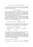
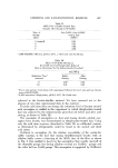
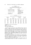
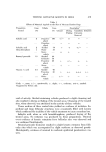
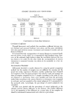
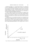
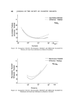
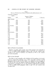
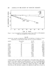
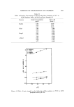
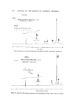
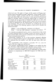
TESTING ANTIACNE AGENTS IN DOGS 475 Preparation Tested Table I Effects of Materials Applied on the Skin of Mexican Hairless Dogs _ Conc. Vehicle Conc. Description • (%) (%) Cleaner Salicylic acid 3 Propylene 100 q-/- glycol ...... Propylene ... -- glycol Salicylic acid 2 Alcohol 70 q- Hexachlorophene 1 Water 30 ...... Above ß ß ß -4- vehicle Benzoyl peroxide 10 AltohoP' 40 Cellosolve 40 Propylene 20 q- glycol ...... Above ... q- vehicle Vitamin A acid 0.1 Alcohol 70 Propylene 30 q- glycol ...... Above ... +/- vehicle Drying, Flaking Comedone Irritation Extrusion Removal JI- + +/- + -- +/-- q- ++ + +++ +q- aCode: --, none +/--, questionable +, slight q-+, moderate 4'-4-+, marked. •Suggested by Fulton (12). void of activity. Alcohol-containing vehicles produced a slight cleansing and also resulted in drying or flaking of the treated areas. Cleansing of the treated sites, when observed, was attributed to the activity of these vehicles. Tissue sections of these control sites yielded no evidence of irritation. Su- perficial and deep fo]licular structures were consistently filled with keratin which confirmed the inability of the vehicle treatment to cause comedone loss. Salicylic acid, alone or with hexachlorophene, produced drying of the treated areas. No irritation was produced by these preparations. Minimal overt evidence of keratin extrusions from follicular sites was observed and was confirmed histologically. Benzoyl peroxide treatment resulted in slight keratin extrusion from folli- cular sites which was accompanied by slight erythema as observed grossly. Histologically, evidence of minimal to moderate epidermal parakeratosis was seQn.
Purchased for the exclusive use of nofirst nolast (unknown)
From: SCC Media Library & Resource Center (library.scconline.org)