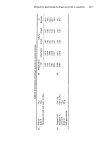
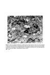
622 J. A. A. Hunter The pink appearance of untanned Caucasoid skin is due to the reddish pigment haemoglobin in the small blood vessels of the superficial dermis. If a sun tan is acquired this pink colour is partly obscured by a brown pigment, melanin, in the overlying epidermis. Melanin is, of course, also responsible for the various shades of brown seen in Negroids. Other hues are produced by the combination of these pigments with the fourth one, carotene, a yellow substance found in the subcutaneous fat and epidermis. There is no blue pigment, and when this colour is seen, it is the result of an optical effect (1). Abnormal skin colour may either be produced by an imbalance in the proportion of these four pigments, as seen for example, in cyanosis, diffuse melanosis and caro- tenaemia, or may be caused by an abnormal pigment (Table II). Table H. Some pigments responsible for abnormal skin colour HaemoglobinSproducts Methaemoglobin Sulphaemoglobin Carboxyhaemoglobin Billrubin and Biliverdin Haemosiderin Metals Gold, Silver Drugs Mepacrine, Clofazimine, Chlorpromazine Tattoo Pigments Although melanin is therefore only one of many pigments, the rest of this paper will be devoted solely to the study of mechanisms involved in pigmentation due to melanin. in emphasising structural aspects an attempt will be made to set the scene of melanin formation and disposal. Later papers will both amplify some of the points mentioned here only briefly and expand the general theme to include discussions on the biochemistry, control and significance of melanin pigmentation. CELLULAR SITE OF MELANIN SYNTHESIS The pioneer work of Bloch (2) indicated that certain branched cells in the epidermis now called melanocytes, contained an oxidising factor (dopa-oxidase) which catalysed the oxidation of the coloufiess dihydroxyphenylalanine (dopa) to an insoluble dense substance, melanin. Improvements in Bloch's dopa technique (3) led the Canadian Masson (4) to propose that the dopa positive melanocyte was the only melanin producing cell in the epidermis. Fitzpatrick, Becker, Lerner and Montgomery (5) modifying Bloch's method by using tyrosine instead of dopa as a substrate, demonstrated the presence of tyrosinase in melanocytes. Most workers today consider tyrosinase and dopa-oxidase to be identical the enzyme catalysing both tyrosine and dopa in the initial steps of melanin formation. Billingham (6) showed that the branches of melano- cytes, seen in the epidermal basal layer, travelled along the intercellular spaces between ordinary epidermal cells (keratinocytes), split frequently, and terminated in the form of 'caps' or 'end buttons' closely applied to the walls of keratinocytes. He felt that melanin granules were manufactured within the melanocyte and passed to the surrounding

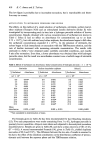
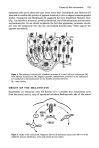
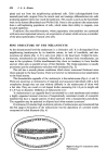
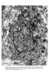
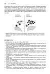
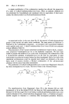
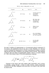
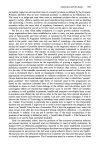
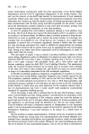
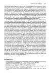
Causes of skin colouration 623 epidermal cells across these end caps. Some years later Cruickshank and Harcourt (7) were able to confirm this process of pigment donation in vitro in elegant cinematographic studies. Fitzpatrick and Breathnach (8) suggested the term 'Epidermal Melanin Unit' (Fig. 1) to describe a structural, as well as functional, use of the melanocyte and surround- ing keratinocytes. Its use should emphasize the fact that pigmentary processes involve not only the melanocyte but also the surrounding keratinocytes, which acquire the pigment secondarily. Fi•m 1. The epidermal melvin unit. Den•itic pro•sses of a basal •11 layer melanocyte (M) wind betw•n keratinocytes (K). Pigment granules, melanosomes, produced in •e melanocyte •e transferred to •e keratinocytes via the den•itic processes (•rows). BL: basal lamina. ORIGIN OF THE MELANOCYTE Experiments on embryonic mice led Rawles (7) to conclude that melanocytes arise from the neural crest (a strip of specialised ectoderm flanking each side of the neural Figure 2. Origin of the melanocyte. Migration (arrow) of embryonic neural crest cells (*) to the epidermis, mucous membranes, retina and central nervous system.
Purchased for the exclusive use of nofirst nolast (unknown)
From: SCC Media Library & Resource Center (library.scconline.org)