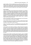
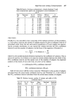
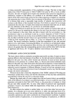
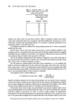
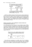
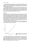
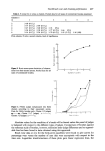
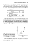
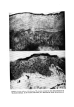
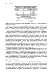
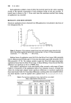
438 R. Marks Table VHI. Results obtained after application of aqueous cream for 14 days using cohesography technique Time (days) n Mean grams force + S.D. 0 7 109'1+49'0 7 8 94.4+34.3 14 5 85'2+ 19'9 Table IX. Cohesography results before and 3 h after application of HA Time (h) Grams force (Mean_+ S.D.) 0 (control) 222 + 69-2 3 157.7 + 64' 3 Table IJf, however, documents one such experiment using an oil in water emulsion in three normal subjects. We have also started to use a method that measures 'point penetrability' (8) and which depends on the measurement of the force required for a rapidly moving needle to move a measured distance into the SC. So far we have found that 'acute hydration' with a water soaked gauze pad results in a drop in the force required of 20-30•o. Both the above techniques utilise important physical properties of the SC as indicators of hydration. The next in vivo technique I want to briefly mention does not appear to be a measure of an important physical property, but is probably a function of many vari- ables. This is the measurement of the electrical properties of the skin. Clar, Her and Sturelle (9) have made significant contributions to this topic and this group claims that low frequency impedance is located almost entirely in the horny layer. They and others (10) have derived complex relationships between impedance and hydration based on the movement of ions in hydrated SC. Certainly, these groups appear to be able to evaluate HAs using this approach although the constraints necessary to obtain consistent results would appear to render the technique awkward for routine use. Our own experience with electrical measurements is very limited and although we believe that the techniques can be useful we intend to evaluate them alongside the methods with which we are more familiar, before making a commitment to their regular use. Another approach is to measure the changes in transepidermal water loss (TEWL) after hydration. The water barrier property of the SC is quite dramatically changed after relatively short hydration. For example, Quattrone et al. (1) report a 20-40• increase in TEWL after 5 min hydration which reverts to normal within a 30 min period. When an 'emollient cream' was used there was a 20• decrease after 1 h and a 10• decrease after 4-6 h. The decrease after the use of an occlusive HA presumably results in hydration of the SC from the movement of H•O into the SC from the living tissue beneath. This same author reviews other workers' experiences employing TEWL. (e) In Vitro TECHNIQUES It is difficult to be enthusiastic concerning in vitro techniques for the evaluation of HA. The SC is so utterly dependent in vitro on the ambient environmental temperature and relative humidity (11) that controlled environments and long periods of equilibration are necessary. Furthermore, the application of topical agents to the in vitro specimen does not accutely emulate the 'in use' situation. The in vitro techniques that have been used include measurements that depend on extensibility and elasticity (12, 13) differential

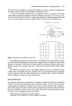
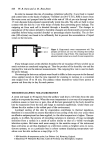
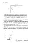
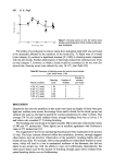
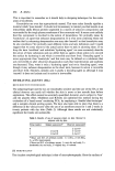
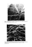
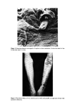
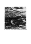
Evaluation of emollients and keratolytics 439 scanning calorimetry (14), gravimetric techniques (15), and photoacoustic spectro- scopy (16). The last of these has recently been adapted for in vivo use and I am told (Pines, personal communication) that the early results are extremely promising. Clearly such techniques may be useful to screen compounds before formulation or to attempt to dissect out the mode of action of HAs but I doubt whether they will have much application to the routine evaluation of these materials. DESCALING AGENTS (DAS) We are in an even worse state for the evaluation of DAs than for HAs and I could begin and end by saying that there are no useful techniques available. I will attempt, however, to summarise briefly our experience so far and indicate what further work, we plan. It seems that the most efficient method to date depends on clinical evaluation. Van Scott and Yu (17) used patients with ichthyotic disorders to investigate the descaling effect of a number of alpha-hydroxy acids and were certainly able to make recommendations on the basis of their clinical observations. Our own studies confirm the usefulness of this approach. Figure 4 shows a patient with a rare and severe form of ichthyosis whose right arm was treated with 6• salicylic acid in white soft paraffin twice daily for 10 days and whose left arm received just the vehicle. Fortunately, such patients are rare and it is just not a practical proposition to use this approach routinely. Clearly, it would be ideal to investigate DAs on normal human (or less ideally on animal) skin in vivo or in vitro. The real problem is knowing which parameter to measure. I have implied by my use of the term DA that the best kind of measurement would be on the rate of desquamation. Unfortunately, this is a most difficult measurement to make. We have examined the number of corneocytes liberated after a standardised 'scrub' stimulus to the skin surface using a specially constructed apparatus (18). We have not as yet, however, satisfactorily demonstrated increased numbers of cells liberated from a DA treated site. It may be that this technique is not sufficiently sensitive to pick up differences in normal skin. It might also be mandatory to use abnormally keratinised skin to demon- strate the effect. It is possible, however, to demonstrate the action of DAs on normal SC by either SEM (19) using SSBs, or by actually counting the cell strata within the SC. This last technique is quite promising but unfortunately does necessitate biopsy. The tissue is sectioned on a cryostat and the SC demonstrated by the McKenzie technique (20). Figure 5 is an SEM of SC taken after 10 days use of 6• salicylic acid in white soft paraffin and Fig. 6 shows cryostat sectioned skin treated by the same material compared to a vehicle treated specimen. Other workers (21) have also demonstrated this SC thinning effect of DAs although the way they accomplish this remains mysterious. In vitro testing of DAs has been totally unsuccessful in our hands. We have applied keratolytics to SSBs themselves and demonstrated no change in surface contour. We plan many more studies with DAs both in vitro and in vivo using measures of the rate of cell loss and the tensile properties of the SC. CONCLUSION We are on the threshold of an exciting era in dermatology and cosmetic science. I believe that in the not too distant future there will be accurate and convenient techniques for the evaluation and measurement of many of the skin's properties and functions. This cer- tainly appears to be the case for HAs and I am certain that with perseverance the same will shortly be true for DAs as well.
Purchased for the exclusive use of nofirst nolast (unknown)
From: SCC Media Library & Resource Center (library.scconline.org)