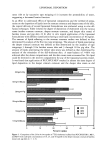
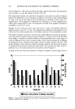
86 JOURNAL OF THE SOCIETY OF COSMETIC CHEMISTS Since either anti-oxidants or anti-inflammatory agents alone provide protection, it is likely that a combination of the two would have greater effectiveness. In this report, we describe the protective effect in hairless mice of the combination against UV radiation- induced chronic skin damage. The degree of protection is quantified using both histo- logical and visible perception methods. MATERIALS AND METHODS ANIMALS Female albino hairless Skh:HR-1 mice were obtained from the Temple University Health Sciences Center, Philadelphia, PA. They were housed five to a cage in a room with controlled temperature and humidity and with a 12-hour light/darkness cycle. They were given a standard diet and water ad libitum. Mice were approximately 10 weeks old at the start of experimental work. At the end of experimental work, they were sacrificed by CO 2 asphyxiation. IRRADIATION AND TOPICAL TREATMENT The procedure for irradiation of the dorsal skin of mice with UVB or UVA radiation has been described previously (6,8). Briefly, mice were irradiated individually under a bank of four Westinghouse FS-40 suniamps (UVB radiation, peak of emission near 315 nm) or four General Electric F-40 black lights (UVA radiation, no detectable emission below 340 nm). The output of the sources was monitored with an International Light (New- buryport, MA) model IL1350 radiometer with model SED240 (UVB) and SED015 (UVA) sensors. Mice were irradiated three times weekly (Monday, Wednesday, and Friday) with 30 mJ/cm 2 UVB radiation per exposure (approximately one half the mouse MED) or five times weekly (Monday-Friday) with 15 J/cm 2 UVA radiation per expo- sure. For mice receiving topical treatment with anti-inflammatory agents and/or anti- oxidants, the dorsal skin of the mice (n = 10 per group neck to tail area) was treated with 0.1 ml of test material solution two hours prior to each UV radiation exposure. Solutions were w/v% (pH 6-7) in an ethanol:propylene glycol:water vehicle [typically 2' 1' 1 (v:v:v)], the ratio of the components varying somewhat, depending upon the solubility of the particular test material. The test solution was delivered to the skin using a Pipetman © (Rainin Instrument Co., Woburn, MA) and spread evenly over the dorsal skin with the flat edge of the disposable piper tip. VISIBLE SKIN GRADING Skin wrinkling (UVB radiation-induced event) and skin sagging (UVA radiation- induced event) in hairless mice were assessed as described previously (6-8). Briefly, wrinkling and sagging were evaluated using grading scales (0-3, with half-grade in- crements), where 0 is normal and 3 is the maximum visible skin change observed in our work (6). Skin lesions were diagnosed as tumors if they were circular, red, raised, and greater than 1 mm in diameter. These lesions were counted.

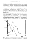
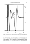
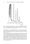
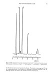
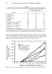
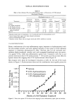
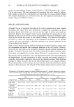
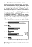
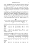
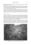
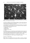
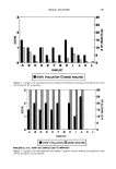
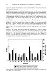
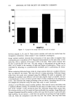
TOPICAL PHOTOPROTECTION 87 HISTOLOGY The histological methods and grading scales were those used previously (6,7,9). Briefly, biopsies (2 x 10 mm) from visibly non-tumor-bearing dorsal skin were taken at sacrifice (CO 2 asphyxiation), fixed in 10% buffered formalin, embedded in paraffin, and sec- tioned at 6 to 10 microns. Sections were then stained with a variety of stains. The grading scales were based on our observations of the sequence of histological events that occur in the mouse, 0 representing the histological observations on normal 10- to 12-week-old mice, and the upper end of the scales being the maximum observed change in the parameters in previous work (6). The histological parameters and the correspond- ing stains and grading scales (half-grade increments) follow: epidermal thickness (H&E stain), 0-3 scale glycosaminoglycan content (Mowry stain), 0-4 scale dermal cellu- larity (H&E stain), 0-3 scale elastosis (Luna stain), 0-5 scale and collagen damage (Van Gieson stain), 0-5 scale. The scores for all parameters were summed to arrive at a histological damage index, a convenient way to express histological data in a single number. STATISTICS The Student t-test was used to determine the statistical significance of differences among treatment groups. RESULTS AND CONCLUSIONS UVB PHOTOPROTECTION Binary combinations of an anti-inflammatory agent (naproxen, ibuprofen, or hydro- cortisone) with an anti-oxidant (alpha-tocopherol, ascorbic acid, or 2,4-hexadien-l-ol) were applied topically to mice prior to each UVB radiation exposure to determine photoprotective effect. The combinations were more effective against mouse skin wrin- kling than were the individual test materials (Table I). The level of protection by the combination appears to be additive of the individual component effects. A plot of the data for a typical example, 0.5 % hydrocortisone q- 5 % alpha-tocopherol, is presented in Figure 1. Noteworthy is the delay in onset of wrinkling by the combi- nation. There was no delay for the individual components. Skin biopsies were taken for histological evaluation at week 20, the end of the study shown in Figure 1. The histological results parallel the visible skin-wrinkling data i.e., the combination was overall more protective against alterations than were the individual components (Table II). As expected, dermal cellularity scores were reduced for the treatments involving an anti-inflammatory agent. Of particular interest are the collagen histological scores, which correspond well with the observed skin-wrinkling grades. Other data from our lab (2,9,10) also suggest a similar association. The combination treatment groups were stopped before a good assessment of tumor development could be made. However, most vehicle control mice had at least one visible
Purchased for the exclusive use of nofirst nolast (unknown)
From: SCC Media Library & Resource Center (library.scconline.org)