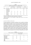
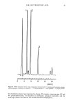
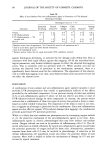
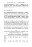
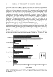
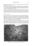
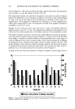
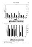
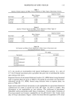
90 JOURNAL OF THE SOCIETY OF COSMETIC CHEMISTS Table III Effect of Anti-Oxidant Plus Anti-Inflammatory Agent in Prevention of UVA-Induced Histological Changes Histological scores (n = 5) a Epidermal Dermal Collagen GAG Treatment thickening cellularity b damage increase Elastosis b Damage index c 0.5% 0 I 1.5] 0.2 0.1 3.1 0.5% Hydrocortisone q- 5% ascorbic acid 0.2
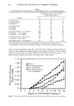
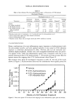
Hydrocortisone 1.5 0.4 2.4 ] No UV 0 0.6 ] 0.1 0.4 ] 6.8] 4'91 4.5 1.1] Baseline scores (start of experiment, 10-12-week-old mice) for all parameters are O. Sum of scores from upper and lower dermal evaluations. Sum of all histological scores. Brackets enclose values that are equal statistically (95% confidence interval). against histological alterations, as indicated by the damage index (Table III). This is consistent with their equal efficacy against skin sagging. Of all the individual histo- logical parameters, only dermal cellularity appears to reflect the observed skin-sagging scores. This is consistent with our previous work (7). While ascorbic acid did not increase the observed level of protection in the combination treatment, there is a significantly lower elastosis score for the combination. The importance of this observa- tion to visible skin sagging is not clear, since hydrocortisone alone was protective yet did not affect the elastosis score. DISCUSSION A combination of anti-oxidant and anti-inflammatory agent applied topically to mice provides UVB photoprotection that overall is approximately additive of the effects provided by the individual components. In our previous testing of either anti-oxidants (2,3) or anti-inflammatory agents (7) alone, we did not observe any delay in onset of UVB radiation-induced damage by these compounds. In contrast, the present results indicate that a combination of these two types of actives does provide a delay in onset, based on visible wrinkle evaluations. The importance of the delay in onset is not clear, but it suggests an advantage in protection for the combination. Since anti-inflammatory agents are also effective against UVA-induced damage (skin sagging), the combination provides good broad UV spectrum protection. While it is likely that anti-oxidants are photoprotective by scavenging oxygen radicals (1-3), the protective mechanism of the anti-inflammatory agents is not clear. Anti- inflammatory agents in chronic photodamaged skin may have indirect anti-oxidant action. Oxygen radicals can arise as by-products of inflammatory cells (11). The ob- served reductions in dermal cellularity by topical anti-inflammatory agents would lessen the potential generation of oxygen radicals by these cells. Alternatively, proteolytic enzymes from these cells (12) may be involved in photodamage. A reduction in the dermal inflammatory cell population would also reduce the potential release of such enzymes. More work is needed to define the mechanisms of damage by UV and pro- tection by topical treatment.

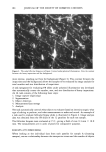
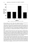
TOPICAL PHOTOPROTECTION 91 Our histological data indicate a high level of elastosis with both UVB and UVA radiation exposures. However, our results here and elsewhere (2) do not reveal an obvious association between the effectiveness against elastosis and the efficacy of a topical treatment against visible skin changes and other histological parameters. While elastosis is a useful marker for identifying photodamaged skin, it is not clear how it relates to other photodamage events. Further effort is necessary to define the association among these events. We believe that the results observed here are relevant to human photoprotection. In human testing, both anti-oxidants and anti-inflammatory agents have been shown to be effective against acute skin photodamage (13-18). Tocopherol is protective against lipid peroxidation (13), while ascorbic acid reduces the severity of UV-induced erythema (14). Several anti-inflammatory agents also inhibit UV-induced erythema (15-18). Addition- ally, we have observed that both tocopherol and hydrocortisone inhibit the UV induc- tion of human epidermal ornithine decarboxylase and that acute photoprotection pre- dicts chronic photoprotection (manuscripts in preparation). We therefore anticipate that the acute observations on humans will translate into chronic benefits for anti-oxidants and anti-inflammatory agents, used either alone or in combination. ACKNOWLEDGMENTS We gratefully acknowledge the technical assistance of James F. McBride, Rebecca A. Runyan, Larry F. Patrick, Mark J. Benzinger, and Jayne L. Ritter. REFERENCES (1) H. S. Black, Potential involvement of free radical reactions in ultraviolet light-mediated cutaneous damage, Photochem. Photobid., 46, 213-221 (1987). (2) D. L. Bissett, R. Chatterjee, and D. P. Hannon, Photoprotective effect of superoxide-scavenging anti-oxidants against ultraviolet radiation-induced chronic skin damage in the hairless mouse, Pho- todermatol. Photoimmunol. Photomed., 7, 56-62 (1990). (3) D. L. Bissett, S. Majeti, J. L. Fu, J. F. McBride, and W. E. Wyder, Protective effect of topically applied conjugated hexadienes against ultraviolet radiation-induced chronic skin damage in the hair- less mouse, Photodermatol. Photoimmunol. Photomed., 7, 63-67 (1990). (4) D. L. Bissett, R. Chatterjee, and D. P. Hannon, Chronic ultraviolet radiation-induced increase in skin iron and the photoprotective effect of topically applied iron chelators, Photochem. Photobiol., 54, 215-223 (1991). (5) L. H. Kligman, F. J. Akin, and A.M. Kligman, The contributions of UVA and UVB to connective tissue damage in hairless mice, J. Invest. Dermatol., 84, 272-276 (1985). (6) D. L. Bissett, D. P. Hannon, and T. V. Orr, An animal model of solar-aged skin: Histological, physical, and visible changes in UV-irradiated hairless mouse skin, Photochem. Photobiol., 46, 367-378 (1987). (7) D. L. Bissett, R. Chatterjee, and D. P. Hannon, Photoprotective effect of topical anti-inflammatory agents against ultraviolet radiation-induced chronic skin damage in the hairless mouse, Photodermatol. Photoimmunol. Photomed., 7, 153-158 (1990). (8) D. L. Bissett, G. G. Hillebrand, and D. P. Hannon, The hairless mouse as a model of skin photo- aging: Its use to evaluate photoprotective materials, Photodermatol., 6, 228-233 (1989). (9) D. L. Bissett, D. P. Hannon, and T. V. Orr, Wavelength dependence of histological, physical, and visible changes in chronically UV-irradiated hairless mouse skin, Photochem. Photobid., 50, 763-769 (1989). (10) R. Chatterjee, M. J. Benzinger, J. L. Ritter, and D. L. Bissett, Chronic ultraviolet B radiation- induced biochemical changes in the skin of hairless mice, Photochem. Photobid., 51, 91-97 (1990).
Purchased for the exclusive use of nofirst nolast (unknown)
From: SCC Media Library & Resource Center (library.scconline.org)