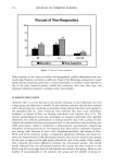
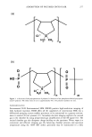
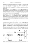
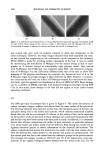
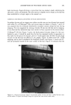
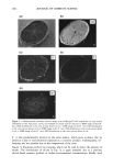
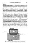
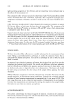
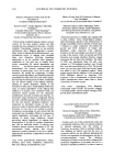
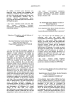
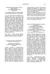
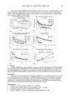
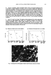
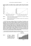
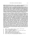
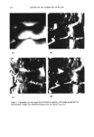
ADSORPTION OF POLYMER ONTO HAIR 283 light microscopy. Figure 6b shows a typical hair that was studied, clearly exhibiting the epicuticle, cortex, and medulla. The cross section is slightly oval in shape, as the hair has been embedded at a slight angle to the normal cut. ELEMENTAL AND MOLECULAR MAPPING OF HAIR CROSS SECTIONS Secondary electron and ion images were taken over the cross section of many hairs treated with either N- or P-Merquat©-100, and typical maps are shown in Figures 7 and 8. It was assumed that the elemental distribution remained constant along the hair during imaging. This was supported by etching a further 20 nm into the hair and re-imaging. It was observed that elemental maps were similar to those acquired on the initial surface. N-Merquat©-100 hair. Figure 7a gives the backscattered electron image of a hair em- bedded in resin. It should be noted that the hair has remained similar in appearance to that taken by the optical microscope, and so it would appear that the effect of ultra-high vacuum on the hair has not affected it too greatly. Figure 7b shows how CN- (26D) mapping can be used to emphasize the biological matrix, and Figure 7c shows how O- sites are primarily located in the outer surface of the hair. Figure 7d indicates that the (a) (b) Figure 6. a: Optical microscope image of microtomed surface of resin and hair to allow flat hair cross- section studies. b: Optical microscope image of a typical hair cross section under study.

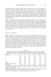
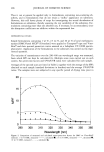
284 JOURNAL OF COSMETIC SCIENCE (a) (c) (b) (d) CN- (e) Figure 7. a: Backscattered secondary electron image of an N-Merquat©-100-treated hair in cross section embedded in resin. Epicuticle, cortex, and medulla are clearly seen for this hair. b: SIMS image of the CN- (m/z 26D) distribution in the cross section shown in (a). c: SIMS image of the O- (m/z 16D) distribution in the cross section shown in (a). d: SIMS image of the F- (m/z 19D) distribution in the cross section shown in (a). e: SIMS image of the Si + (m/z 28D) distribution in the cross section shown in (a). F- is also predominantly located on the outer surface, which gives evidence that its presence is due to environmental exposure or a cosmetic product. Unfortunately, CI- imaging was not possible due to the composition of the resin. Figure 7e illustrates positive ion imaging, which can be used to detect the presence of metals. The distribution of silicon in Fig 7e is again probably due to a previous silicon-based cosmetic product or surface environmental contamination. Ideally, both
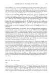
Purchased for the exclusive use of nofirst nolast (unknown)
From: SCC Media Library & Resource Center (library.scconline.org)