200 JOURNAL OF COSMETIC SCIENCE Lymphatic and venous circulation in the tissue are impaired, and exchanges between the blood, the lymph, and the tissues are decreased (2,3). This may contribute to the damage of the fibroelastic strands. A vicious cycle ensues, in which enlarged fat 1obules increase the hypodermal pressure, further damaging the vessels and degrading the fibers (colla- gen and elastin fibers). Fat lobules are squeezed upward upon pinching the skin, while their belt of fibrosclerotic strands acts like shrouds bound to the deepest fascia (4). The skin on the thigh in women is easily deformed because the dermis is thin and the collagen bundles and the elastic network are vertically oriented, leading to the "orange peel" effect when the fibers are damaged. As the condition worsens, the dimpled aspect of the skin occurs spontaneously, without even pinching the skin. This study evaluated the effect of the association of three active ingredients present in a cosmetic product: caffeine, ruscogenine extract, and retinol. The evaluation of the test product was performed using an original combination of objective methods, which had not yet been used: ß Profilometric analysis of the skin's macrorelief of the external face of the thighs performed on digital images of pinched thighs ß 3D ultrasound imaging (evaluation of the dermal and hypodermal structures) ß Cutometry (evaluation of the mechanical properties of the skin of the thighs) ß Laser Doppler flowmerry (evaluation of the cutaneous microcirculation) MATERIAL AND METHODS PANELISTS Forty-six (46) healthy female volunteers, minimum 18 years of age, with normal skin were selected. They presented with a moderate level of cellulite in the thighs, and their weight/height ratio [body mass index (BMI) = weight/height] was between 20 and 25. All volunteers gave their informed written consent before beginning the test. STUDY DESIGN The format was a randomized, double-blind, placebo-controlled study. The study lasted three months (from the beginning of March until the end of May, 1999), the products were applied twice a day for three months (one thigh treated with the product and the other with the placebo). Measurements were taken before the first application, and then after 28 days, 56 days, and 84 days of application. PRODUCTS The complete product contained retinol, caffeine, ruscogenine extract, and alcohol. The placebo did not contain retinol, caffeine and ruscogenine extract (the alcohol was main- tained to give the same cosmetic perception on application). APPLICATION OF PRODUCTS The volunteers applied both products regularly and uniformly over the entire thigh by circular massage. Products were applied twice daily, under in-use conditions.

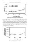
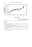
ANTI-CELLULITE ACTIVITY 201 POSITIONING OF SITES Measuring sites on the thigh were marked with indelible ink after visual positioning at the summit of the cellulite bulge on the exterior lateral aspect of the thigh, at the same height from the ground for each thigh. EVALUATION OF EFFECTIVENESS Effectiveness was evaluated by the measurement of different parameters and their evo- lution: © Macro-relief of the skin ("orange peel" effect) © Structure of the dermis and hypodermis © Mechanical characteristics of the skin © Flowmerry of the skin perfusion The following measuring devices were used: 1. Macrorelief of the skin: Profilometry on digital imaging of pinched thighs The technique consists of acquiring macroscopic photographs of an area on the external lateral aspect of the thighs (5). In order to accentuate the unevenness of the skin's surface caused by cellulite, a localized controlled pinching was applied to the thigh using a compression system under standardized conditions (for each volunteer, the same pressure was applied throughout the study, around 200 g/cn-•2). An incident lighting system was used to accentuate the relief and the skin dimple. All images were acquired with a CCD-RGB type XC-711/711P camera (Sony) equipped with a 60-ram focal length lens (Nikon). Digital images were analyzed using a dedicated software package (Visilog 5.00-NOESIS). Image analysis consisted of measuring the macrorelief of the skin sur- face. The zone under study corresponded to 15 joinrive profiles (1 pixel width x 650 pixels length) corresponding to a skin surface area of 390 mm 2. The conventional Rz roughness parameter frequently used to describe cutaneous relief was used. In our case, this parameter was estimated from the slope a• between two contiguous pixels (calculated from gray-level values). 2. Structure of the dermis and hypodermis: 3D ultrasound imaging Significant parameters quantifying cellulite had been determined during a previous study (6). So,ograph. The imaging equipment used was a sonograph from Esaote Company (AU4- AU5 model) provided with a high-frequency 20-MHz probe with low penetration (19-ram maximum) intended for the imagery of the skin, in particular of dermis and hypodermis. Acqaisitio, system a,a/#ata processi,g. The images delivered by the sonograph were taken on the external lateral surface of the thighs via the video exit of the apparatus by a dedicated workstation (I6 3.3, I6DP, Paris, France). Three-a/ime,sio,a/se,sor. The sonograph delivered an image in real time of the studied area. The probe was then moved in a predefined motion, allowing a digitalized sequence of images representative of the area covered to be generated. Thus, to analyze the parameters of cellulite, it was preferable to acquire sequences of
Purchased for the exclusive use of nofirst nolast (unknown)
From: SCC Media Library & Resource Center (library.scconline.org)