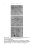
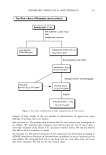
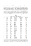
INHIBITORY EFFECTS OF R. A/JORI EXTRACTS 135 INHIBITION OF MELANOGENESIS B-16 melanoma cells were placed in a 25-ml T-flask at a density of 1 x 10⁵ cells/flask and cultured at 37øC in Dulbecco's Modified Eagle's Medium (DMEM, Sigma) con- taining 4.5 g/1 oFglucose, 10% (v/v) Fetal bovine serum (FBS), and 1% (v/v) antibiotic- antimycotic (Gibco, Auckland, N.Z.). AFter 24 hours of cultivation, we replaced the medium with new DMEM medium containing R. •orJ extracts of various concentra- tions. AFter five days, we washed the cells with phosphate-buffered saline (PBS) and collected the cells by trypsinization and centrifugation. We separated melanin From the pellet of the cells using 5% (w/v) trichloroacetic acid, dissolved the melanin in 1N NaOH solution, and checked the melanin content by absorbency at 475 nm. EVALUATION OF R. MORI EXTRACTS ON TYROSINE SYNTESIS IN B-16 MELANOMA CELLS (TYROSINASE ZYMOGRAPHY) Analysis of tyrosinase synthesis was performed by the modified method of Imokawa and Mishima (5). Detergent-solubilized cell extracts were subjected to SDS gel electropho- resis as Follows: Cell extracts (2 mg/ml protein) were electrophoresed on 10% (w/v) polyacrylamide gels. Total protein was measured by protein assay kit (Bio-Rad Labora- tories, California). AFter electrophoresis, the gel was placed in renaturation buffer [50 mM Tris-HCl (pH 8.0) and 2.5% (v/v) triton X-100] at room temperature For one hour. The gel was then incubated in developing buffer [0.1 M sodium phosphate (pH 6.8), 0.2% (w/v) L-DOPA] at 37øC For Four hours. Upon visualization of the tyrosinase bands, the gel was removed and dried, and the relative amount of tyrosinase band in each lane was quantified. EVALUATION OF R. MORI EXTRACTS ON TYROSINASE GENE EXPRESSION IN B-16 MELANOMA CELLS (RT-PCR) Total RNA was prepared using RNA PLUS TM (Quantum, Quevec, MW) From B-16 melanoma cells. Five micrograms per milliliter of total RNA was reverse transcribed by incubating the sample For one hour at 42øC in 25 pl of reaction mixture containing 200 U of MMLV (Moloney murine leukemia virus reverse transcriptase, Promega, Madison, WI) 1 pl of 100 pmol sequence specific primer dNTP (dATP, dCTP, dGTP, dTTP, Promega) and 1X buffer (50 mM Tris-Hcl, 75 mM KCI, 3 mM MgCI• and 10 mM DDT). Ten milliliters of RT reaction mixture was added to 40 pl of PCR mixture containing 1X PCR buffer (50 mM KC1, 10 mM, Tris-HC1 (pH 9.0), 1.5 mM, MgCI•, and 0.1% Triton X-100) 1 pl of 100 pmol Forward and reverse primer 5 pl of 2.5 mM each dNTP 2 pl of 25 mM MgCI• and Five units of Tag DNA polymerase (Promega). Amplification was performed at 33 cycles at 94øC For 30 sec, 50øC For 30 sec, and 72øC For 50 sec with the Gene AMP PCR system 2400 (Perkin Elmer, Oak Brook, IL). Two microliters of loading dye were added to 10 pl of amplification products, and the mixture was analyzed by 2% Agarose (Sigma) gel electrophoresis. EVALUATION OF R. RIORI EXTRACTS ACTIVITY ON MELANOGENESIS IN ANIMAL TEST R. mori extracts were dissolved at a final concentration, 1%, 5% (v/v) in dissolving solution (butylene glycol: H20 = 50:50). This solution was topically applied to sepa-

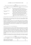
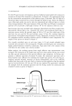
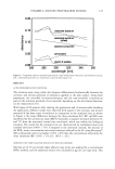
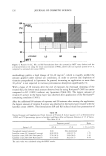
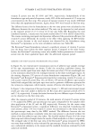
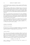
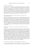
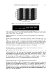
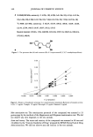
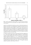
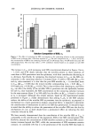
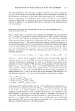
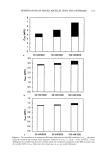
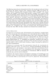
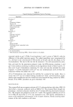
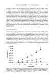
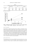
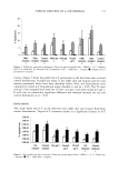
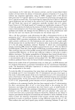
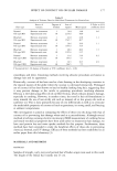
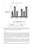
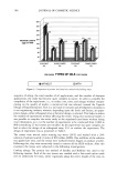
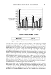
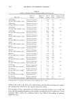
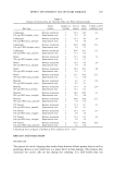
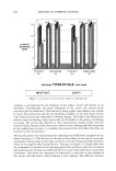
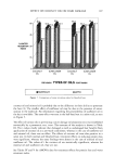
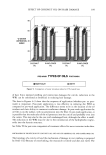
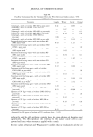
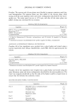
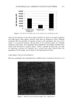
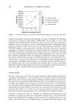
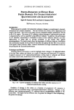
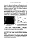
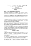
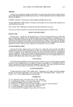
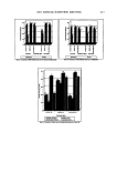
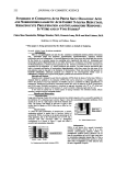
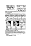
136 JOURNAL OF COSMETIC SCIENCE rated areas on the back skin of guinea pigs (n = 9) for two days (200 pl application, twice/day) before UVB radiation, while the dissolving solution alone was applied to the other area as a control. These areas were then irradiated once with 1,350 mJ/cm 2 using a UVB lamp (Vilber Loutmat, Marine La Vallee, France). After two weeks, the UV- irradiated site was stained by the Fontana-Masson staining method. SEPARATION AND IDENTIFICATION OF AN ACTIVE COMPOUND Separation. We extracted dried young twigs with 70% ethanol solution, using a vacuum rotary evaporator to concentrate the extract to dryness. To isolate the tyrosinase inhibitor from ethanol extract, we purified the extract through solvent fractionation, silica column chromatography, and Prep-LC. The ethanol extracts were dissolved in ethyl acetate, and then the residue was crystallized from CHC13. We purified the solid using silica chro- matography (Merck 200-400) and finally isolated the tyrosinase inhibitor. Identification. We crystallized the isolated tyrosinase inhibitor from ether benzene to yield pale yellowish prisms. The compound was identified by infrared (IR) spectroscopy, mass chromatography, and nuclear magnetic resonance (NMR). RESULTS INHIBITION OF TYROSINASE ACTIVITY AND MELANIN SYNTHESIS R. mori extracts were selected as potent tyrosinase inhibitors through our screening methods. We checked the extracts' inhibition activity on tyrosinase and melanin syn- thesis by changing the concentration (10, 20, 50, and 100 pg/ml). The extracts showed high tyrosinase inhibition activity. Melanin synthesis was also inhibited by R. mori extracts at a concentration of 50 pg/ml. The extracts showed no cytotoxicity. See Table I. EFFECT OF R. MORI EXTRACTS ON TYROSINE SYNTHESIS AND GENE EXPRESSION To examine the inhibitory mechanism of R. mori extracts on melanogenesis, we did the zymography for tyrosinase content in B-16 melanoma cells and RT-PCR for tyrosinase gene expression. Tyrosinase zymography showed that R. mori extracts at a concentration of 50-100 pg/ml did not lessen the tyrosinase synthesis (the band intensity does not change) (Figure 1). We also checked whether R. mori extracts inhibit the tyrosinase gene level by using RT-PCR. Figure 2 shows that the tyrosinase gene level was not changed by the treatment of R. mori extracts. These two results mean that R. mori extracts inhibit only tyrosinase activity, not tyrosinase synthesis and tyrosinase gene expression. Table I Inhibitory Effects of R. mori Extracts on Tyrosinase and Melanin Synthesis R. mori extracts (pg/ml) Tyrosinase inhibition (%) Inhibition of melanin synthesis (%) 10 56.0 13.5 20 65.8 18.5 50 89.5 36.5 100 97.8 65.3
Purchased for the exclusive use of nofirst nolast (unknown)
From: SCC Media Library & Resource Center (library.scconline.org)